Atezolizumab: Classification, History, Mechanism of Action, Safety and Pharmacovigilance
- Atezolizumab: Classification, History, Mechanism of Action, Safety and Pharmacovigilance
- Multidimensional classification
- PD-1/PD-L1 biology
- Development and regulatory evolution
- Biomarker and treatment-context interpretation
- Immune-mediated adverse reactions
- Infusion reactions and hypersensitivity
- Pharmacokinetic persistence and delayed events
- Pharmacovigilance case assessment
- Signal detection and aggregate review
- Periodic benefit-risk evaluation
- Risk management and operational controls
- Potential failure modes
- Inspection and governance perspective
- Practical checklist
- Key Takeaways
- References
- Regulatory Note
Atezolizumab is a monoclonal antibody that blocks programmed death-ligand 1 (PD-L1), an inhibitory immune-checkpoint ligand expressed by tumour cells and by immune cells within the tumour microenvironment. Its therapeutic purpose is not to kill a cancer cell directly. Instead, it removes one of the molecular inhibitory signals that can suppress cytotoxic T-cell activity, thereby allowing an existing or newly generated antitumour immune response to function more effectively.
This mechanism changes the logic of pharmacovigilance. With a cytotoxic medicine, an adverse effect is often sought in tissues that are directly exposed to a damaging pharmacological action. With checkpoint inhibition, the relevant mechanism is loss of immune restraint. Almost any organ can therefore become a site of inflammatory injury, and the event may begin during treatment or after treatment has stopped. The same symptoms can also arise from infection, tumour progression, paraneoplastic disease, radiotherapy or concomitant antineoplastic therapy. Causality depends on pattern recognition plus exclusion of plausible alternatives, not on timing alone.
Multidimensional classification
| Classification axis | Atezolizumab classification | Scientific or PV significance |
|---|---|---|
| Molecular class | Humanised IgG1 monoclonal antibody | Biological product with long systemic persistence |
| Target | PD-L1 | Blocks an inhibitory ligand rather than directly targeting a tumour-lineage antigen |
| Receptor interactions blocked | PD-L1 with PD-1 and B7.1 | Releases inhibitory signalling affecting T-cell activity and antigen-presenting-cell interactions |
| Fc design | Fc-engineered IgG1 with reduced effector-cell killing intent | Designed to avoid substantial depletion of PD-L1-expressing immune cells through ADCC |
| Functional class | Immune-checkpoint inhibitor | Benefit and toxicity arise from immune activation rather than direct tumour-cell cytotoxicity |
| Therapeutic class | Antineoplastic immunotherapy | Used across multiple tumour types, often in combination regimens |
| Product category | Biological medicinal product | Product identity, presentation and batch remain relevant to traceability and quality events |
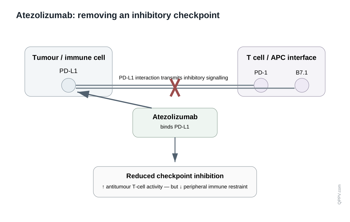
Figure 1. Atezolizumab acts at the PD-L1 checkpoint interface. Blocking PD-L1 interaction with PD-1 and B7.1 removes inhibitory signalling; the therapeutic effect is immune activation rather than direct antibody-mediated tumour-cell killing.
PD-1/PD-L1 biology
T-cell activation requires antigen recognition but is also governed by co-stimulatory and inhibitory signals. PD-1 is an inhibitory receptor expressed on activated T cells and other immune cells. When PD-1 binds PD-L1, intracellular phosphatases are recruited that attenuate signalling from the T-cell receptor and associated co-stimulatory pathways. The result is reduced proliferation, cytokine production and cytotoxic activity.
This inhibitory system is physiologically useful. It limits excessive tissue damage during immune responses and contributes to peripheral tolerance. Tumours can exploit the same pathway by expressing PD-L1 themselves or by existing within inflammatory microenvironments where PD-L1 is induced on tumour and immune cells. A T cell can therefore recognise tumour antigen yet remain functionally constrained by inhibitory checkpoint signalling.
Atezolizumab binds PD-L1 and prevents its interaction with PD-1. It also blocks PD-L1 interaction with B7.1. PD-L2 remains available to interact with PD-1 because the antibody targets PD-L1 rather than PD-1 itself. This is a mechanistic distinction from PD-1-directed antibodies, although both classes converge on release of PD-1-pathway inhibition.
Why Fc engineering matters
PD-L1 is not restricted to malignant cells. It is also expressed by antigen-presenting cells and other immune populations. If a PD-L1 antibody strongly recruited Fc-mediated cytotoxicity, it could theoretically deplete immune cells that participate in antitumour responses. Atezolizumab's Fc region was therefore engineered to minimise clinically relevant antibody-dependent cellular cytotoxicity against PD-L1-expressing cells.
This illustrates an important principle in therapeutic-antibody design: the variable region determines the checkpoint target, while the Fc can be tuned so that binding changes signalling without turning the bound cell into a target for immune destruction.
Development and regulatory evolution
Atezolizumab emerged from the broader development of immune-checkpoint therapy after experimental and clinical evidence established the PD-1/PD-L1 axis as a reversible mechanism of tumour immune escape. Its regulatory history subsequently expanded across several malignancies and treatment settings, including lung, urothelial and other cancers, with indications changing over time as confirmatory trials, biomarker strategies and combination regimens matured.
Current product information must therefore be used for the exact authorised tumour type, disease stage, biomarker requirement and combination regimen. Historical authorisations should not be assumed to remain current merely because they appear in older trials or publications.
This evolving indication map is itself a PV issue. Atezolizumab may be used as monotherapy, with cytotoxic chemotherapy, with targeted therapy or in perioperative/adjuvant settings. The background incidence of pneumonitis, hepatitis, diarrhoea, cytopenias and constitutional symptoms can differ greatly between these contexts. Aggregate safety evaluation should preserve the regimen rather than treating all exposure as interchangeable.
Biomarker and treatment-context interpretation
PD-L1 expression can be measured on tumour cells, tumour-infiltrating immune cells or both, depending on tumour type, assay and regulatory context. The biomarker is therefore not a universal binary property of a patient. It is an assay-defined observation made on a particular specimen, using a defined scoring algorithm and threshold, in a specific indication.
For pharmacovigilance, this matters most when assessing apparent lack of efficacy, off-label treatment or biomarker-related medication error. A report stating only that a tumour was “PD-L1 positive” may be insufficient if the authorised use depends on a particular assay, cell population or threshold. Specimen date and site can also matter because tumour biology may change over time and differ between primary and metastatic lesions.
Combination therapy changes attribution
Atezolizumab is frequently administered with other antineoplastic therapies. The same clinical syndrome can therefore have multiple plausible causes. Diarrhoea may reflect immune-mediated colitis, chemotherapy, infection or another medicine. Dyspnoea can represent immune-mediated pneumonitis, infection, pulmonary embolism, radiation injury, tumour progression or cardiac disease. Abnormal liver tests can reflect immune-mediated hepatitis, hepatic metastases, sepsis, biliary obstruction or treatment-related hepatotoxicity from another agent.
The PV reviewer should preserve a treatment map containing every systemic anticancer medicine, recent radiotherapy, surgery, transplant history and relevant supportive medication. Atezolizumab cannot be assessed in isolation from that context.
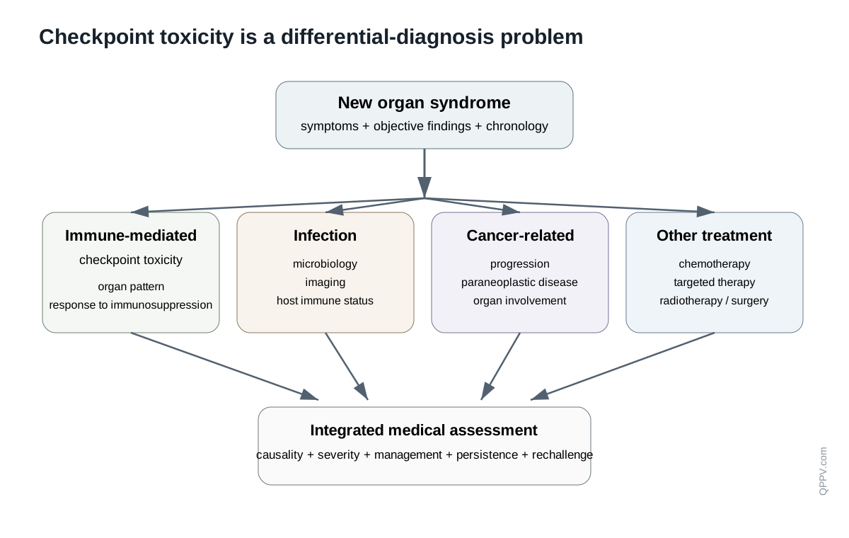
Figure 2. Immune-checkpoint toxicity is a differential-diagnosis problem. Organ inflammation must be interpreted alongside infection, cancer progression, concomitant therapy and other competing causes.
Immune-mediated adverse reactions
Checkpoint inhibition can disturb peripheral immune tolerance. The resulting adverse reactions are often described as immune-mediated because inflammatory pathology occurs in the setting of pharmacological immune activation and may respond to immunosuppression. The label term does not mean that every event with an inflammatory phenotype is proven autoimmune disease.
Pneumonitis
Immune-mediated pneumonitis can present with cough, dyspnoea, hypoxia, fever or radiographic infiltrates. The differential diagnosis is broad in patients with cancer: bacterial or viral pneumonia, tumour progression, lymphangitic spread, pulmonary oedema, radiation pneumonitis and pulmonary embolism may produce overlapping features.
High-value follow-up includes symptom onset, oxygen requirement, CT pattern, infectious testing, bronchoscopy where performed, radiotherapy history, tumour burden, corticosteroid treatment, response, rechallenge and outcome. A spontaneous report of “shortness of breath” cannot support meaningful aggregate interpretation without these contextual elements.
Colitis and diarrhoea
Immune-mediated colitis may range from increased stool frequency to severe inflammatory colitis with bleeding, abdominal pain or complications. Stool studies, imaging, endoscopy and histology can help distinguish immune-mediated disease from infection, chemotherapy-related diarrhoea or other gastrointestinal pathology.
The clinically useful distinction is between a symptom term and an organ diagnosis. “Diarrhoea” may be adequate for initial coding, but aggregate medical review should determine whether the case supports inflammatory colitis, infectious gastroenteritis, treatment-related secretory diarrhoea or another explanation.
Hepatitis
Immune-mediated hepatitis usually presents through abnormal liver biochemistry rather than a pathognomonic symptom pattern. Evaluation should capture ALT, AST, alkaline phosphatase, bilirubin, baseline liver tests, hepatic metastases, viral hepatitis studies, alcohol/metabolic risk, biliary imaging and co-medications. A hepatocellular, cholestatic or mixed biochemical pattern can guide differential diagnosis but does not by itself prove checkpoint causality.
Endocrinopathies
Checkpoint inhibition can cause thyroid dysfunction, hypophysitis, adrenal insufficiency and insulin-deficient diabetes. These events differ from many other adverse reactions because endocrine injury may leave permanent hormone deficiency even after inflammation resolves. Clinical improvement with hormone replacement therefore does not necessarily indicate reversal of the underlying glandular damage.
PV follow-up should collect the relevant hormone profile, timing, imaging where applicable, ketoacidosis or adrenal crisis, long-term replacement requirements and whether immunosuppression was used. Fatigue alone is too nonspecific to support an endocrine conclusion.
Nephritis and renal dysfunction
Immune-mediated nephritis may present as rising creatinine, urinary abnormalities or biopsy-proven interstitial nephritis. Cancer patients also have numerous alternative causes of acute kidney injury, including dehydration, sepsis, contrast exposure, obstruction and nephrotoxic co-medications. Baseline renal function and the temporal relationship to all relevant exposures are therefore essential.
Neurological, cardiac and other rare organ toxicities
Checkpoint therapy can produce uncommon but serious neurological, cardiac, haematological, ocular, dermatological and musculoskeletal inflammatory syndromes. Myocarditis is particularly important because it may progress rapidly and can overlap with myositis or myasthenic features. Neurological syndromes require careful phenotyping because metastases, infection, stroke, metabolic disturbance and paraneoplastic disease are common competing explanations.
The rarity of these events increases the importance of case completeness. Objective tests, specialist diagnoses, imaging, electrophysiology, biopsy and treatment response may materially change signal assessment.
Infusion reactions and hypersensitivity
Infusion-related reactions occur through a different pathway from delayed immune-mediated organ toxicity. Case processing should therefore preserve immediate timing from infusion, vital signs, phenotype, infusion interruption, treatment, recurrence and rechallenge. A reaction beginning minutes into administration should not be merged analytically with delayed autoimmune-like inflammation simply because both occur during immunotherapy.
Pharmacokinetic persistence and delayed events
Atezolizumab has the prolonged disposition expected of a therapeutic IgG antibody. More importantly, checkpoint blockade can initiate immune processes whose clinical consequences persist beyond measurable peak exposure. Adverse events may therefore begin after the last dose or continue after treatment discontinuation.
This disconnect between dosing and immune biology is a central PV principle. A strict short temporal window after the most recent dose can miss plausible treatment-related events.
Pharmacovigilance case assessment
Atezolizumab case assessment should be organised around organ phenotype, competing causes, treatment context and immune-management response. The question is not simply whether the event occurred after exposure. It is whether the clinical pattern is compatible with checkpoint-related inflammation and whether infection, progression, concomitant therapy or another diagnosis provides a stronger explanation.
Event-specific follow-up priorities
| Event | High-value follow-up information |
|---|---|
| Pneumonitis | CT pattern, oxygen need, infection testing, bronchoscopy, radiotherapy, steroids, response, rechallenge |
| Colitis/diarrhoea | Stool frequency, blood, infection studies, imaging/endoscopy/biopsy, IV fluids, steroids/other immunosuppression |
| Hepatitis | Serial ALT/AST/ALP/bilirubin, viral studies, metastases, imaging, co-medications, steroids, recovery |
| Endocrinopathy | Relevant hormones, glucose/ketones, pituitary imaging if applicable, replacement therapy and persistence |
| Nephritis | Baseline/serial creatinine, urinalysis, nephrotoxins, obstruction/infection evaluation, biopsy if performed |
| Myocarditis / myositis | Troponin, ECG, echocardiography/MRI, CK, neuromuscular symptoms, specialist assessment, immunosuppression |
| Neurological syndrome | Detailed phenotype, MRI, CSF, electrophysiology, infection/metastasis evaluation, paraneoplastic work-up |
| Infusion reaction | Onset from infusion, sequence of co-infusions, vital signs, intervention and rechallenge |
Signal detection and aggregate review
Checkpoint-inhibitor signal detection benefits from organ-based medical groupings but should not rely on coding alone. A collection of preferred terms such as diarrhoea, abdominal pain and colitis may represent one inflammatory syndrome, while identical terms in a chemotherapy combination may arise through unrelated mechanisms. Medical review should therefore connect coded events to clinical narratives and objective evidence.
Stratification by tumour type, regimen, line of therapy, route/presentation, recent radiotherapy and relevant baseline comorbidity can reduce confounding. New treatment settings also change the tolerance for long-term toxicity. Permanent hypothyroidism may have a different benefit-risk meaning in a patient receiving curative-intent adjuvant therapy than in heavily pretreated metastatic disease.
Delayed toxicity and post-treatment surveillance
Aggregate methods should allow clinically plausible latency after discontinuation. The immune system does not necessarily return immediately to its pretreatment state when the antibody is cleared. Late endocrine, neurological, pulmonary or other inflammatory events can therefore remain relevant to cumulative evaluation.
Periodic benefit-risk evaluation
Periodic evaluation should integrate exposure by indication and regimen with the complete spectrum of immune-mediated adverse reactions, infusion reactions, serious infection during immunosuppressive management, treatment-related deaths, pregnancy exposure, medication errors, product-quality events and outcomes after rechallenge.
Particular attention should be paid to severe low-frequency syndromes, organ overlap and the consequences of treatment used to manage toxicity. High-dose corticosteroids or additional immunosuppression can themselves generate infection and metabolic complications, creating a second layer of safety assessment.
Risk management and operational controls
Current regional product information governs monitoring, withholding or discontinuation, corticosteroid use and other risk-minimisation instructions. PV systems should support those requirements while preserving the distinction between regulatory instructions and recommended operational practice.
Useful controls include organ-specific targeted follow-up, medical-review algorithms that require competing-cause assessment, structured capture of immunosuppressive treatment, tracking of persistent endocrine replacement and specific conventions for delayed events after discontinuation. Combination-regimen fields should be mandatory for serious events where attribution depends on co-treatment.
Potential failure modes
The following are illustrative scenarios rather than published inspection findings:
- Dyspnoea is coded as pneumonitis without documenting CT findings or excluding infection and progression.
- Diarrhoea is counted as immune-mediated colitis in aggregate review despite positive infectious stool studies.
- A liver injury case omits hepatic metastases and chemotherapy co-exposure.
- Fatigue is labelled hypophysitis without pituitary or hormone evidence.
- A myocarditis case is missed because the event began several weeks after the final dose.
- Serious infection occurring during high-dose corticosteroid treatment is analysed only as a consequence of cancer rather than as part of toxicity management.
- A combination-treatment signal is attributed to atezolizumab without preserving the regimen.
Inspection and governance perspective
An inspector would be interested in whether the system translates the known complexity of checkpoint toxicity into reproducible evidence. Relevant evidence may include organ-specific follow-up forms, medical-review criteria, signal-search strategies, delayed-event rules, treatment-context fields, case reconciliation across oncology and medical-information channels, and documentation of how competing diagnoses were considered.
Effectiveness is demonstrated when the database contains the information required to distinguish immune toxicity from infection and progression. A procedure that says “exclude alternative causes” is not effective if serious cases repeatedly lack imaging, microbiology, concomitant therapy or baseline organ function.
Practical checklist
For an atezolizumab case or aggregate review, confirm:
- tumour type, stage, biomarker context where applicable and treatment intent;
- complete regimen, recent radiotherapy and other relevant anticancer exposure;
- organ-specific objective findings rather than symptom labels alone;
- infection, progression and other competing diagnoses;
- timing relative to all doses, including delayed onset after discontinuation;
- corticosteroid or other immunosuppressive treatment and clinical response;
- persistent sequelae such as endocrine replacement;
- rechallenge and recurrence where clinically relevant;
- exact product/presentation and batch for quality or traceability issues.
Key Takeaways
Atezolizumab is an Fc-engineered humanised IgG1 antibody that blocks PD-L1 interaction with PD-1 and B7.1. Its therapeutic effect is release of inhibitory immune signalling rather than direct tumour-cell killing. That same mechanism produces a distinctive safety problem: inflammatory injury can affect almost any organ and may appear after treatment has ended.
High-quality pharmacovigilance therefore depends on differential diagnosis, complete treatment context and longitudinal follow-up. Pneumonitis, colitis, hepatitis, endocrinopathy, nephritis, neurological and cardiac syndromes should be evaluated using objective organ-specific evidence and active exclusion of infection, tumour progression and co-treatment toxicity.
References
- European Medicines Agency. Atezolizumab: EPAR and current product information. Procedural information last updated 21 July 2026. https://www.ema.europa.eu/en/medicines/human/EPAR/tecentriq
- U.S. Food and Drug Administration. Atezolizumab prescribing information. Current label, 2025. https://www.accessdata.fda.gov/drugsatfda_docs/label/2025/761034s059lbl.pdf
- Herbst RS, Soria JC, Kowanetz M, et al. Predictive correlates of response to the anti-PD-L1 antibody MPDL3280A in cancer patients. Nature. 2014;515:563-567. doi:10.1038/nature14011.
- Fehrenbacher L, Spira A, Ballinger M, et al. Atezolizumab versus docetaxel for patients with previously treated non-small-cell lung cancer (POPLAR). Lancet. 2016;387:1837-1846. doi:10.1016/S0140-6736(16)00587-0.
- Rittmeyer A, Barlesi F, Waterkamp D, et al. Atezolizumab versus docetaxel in patients with previously treated non-small-cell lung cancer (OAK). Lancet. 2017;389:255-265. doi:10.1016/S0140-6736(16)32517-X.
Regulatory Note
Authorised tumour types, biomarker requirements, combinations, dosing schedules and toxicity-management instructions vary by jurisdiction and change as regulatory procedures mature. Historical trial indications should not be treated as current authorisations without checking current product information. Regulatory statements were checked against current EMA and FDA sources available in September 2026. Operational recommendations are pharmacovigilance practice unless explicitly identified as regulatory requirements.