Bevacizumab: Classification, History, Mechanism of Action, Safety and Pharmacovigilance
- Bevacizumab: Classification, History, Mechanism of Action, Safety and Pharmacovigilance
- Multidimensional classification
- Physiological and tumour angiogenesis
- Discovery and development history
- Detailed mechanism of action
- Pharmacokinetics and exposure
- Major safety risks
- Gastrointestinal perforation and fistulae
- Wound-healing complications
- Haemorrhage
- Arterial and venous thromboembolism
- Hypertension
- Proteinuria and renal injury
- Posterior reversible encephalopathy syndrome
- Congestive heart failure
- Ovarian failure, pregnancy and fetal risk
- Infusion reactions and hypersensitivity
- Pharmacovigilance architecture
- Event-specific assessment
- Biosimilars and traceability
- Risk minimisation and effectiveness
- Inspection and governance perspective
- Practical checklist
- Key takeaways
- References
- Regulatory Note
Bevacizumab is the archetypal anti-angiogenic monoclonal antibody. Rather than binding a receptor on a tumour cell, it neutralises vascular endothelial growth factor A (VEGF-A), a soluble and matrix-associated ligand used by tumours and normal tissues to regulate blood-vessel growth, endothelial survival, permeability and repair. This extracellular ligand-neutralisation strategy distinguishes bevacizumab from antibodies directed at tumour-cell antigens and from small molecules that inhibit the intracellular kinase domains of VEGF receptors.
The same pathway explains both benefit and harm. Reducing VEGF-A signalling can limit tumour vascularisation and alter abnormal tumour vessels, but physiological VEGF activity supports endothelial integrity, nitric-oxide-dependent vascular function, glomerular filtration, wound repair, ovarian function and fetal development. Hypertension, proteinuria, bleeding, arterial and venous thromboembolism, gastrointestinal perforation, fistulae, impaired wound healing, posterior reversible encephalopathy syndrome (PRES), ovarian failure and embryo-fetal harm are therefore not an arbitrary collection of toxicities. They form a mechanism-linked safety landscape modified by tumour site, prior surgery, radiation, concomitant chemotherapy and patient vascular reserve.
This article uses the European reference product Avastin as the regulatory anchor. Authorised uses, regimens and precautions are product- and jurisdiction-specific. It separately addresses the widespread off-label intravitreal use of repackaged bevacizumab because that setting introduces different doses, routes, compounding chains and ocular risks; it must not be merged uncritically with authorised intravenous oncology exposure.
Multidimensional classification
| Axis | Bevacizumab classification | Scientific and PV significance |
|---|---|---|
| Molecular format | Monoclonal antibody; IgG1 kappa | Long systemic persistence and Fc-containing biological molecule |
| Species engineering | Humanised antibody | Predominantly human framework with murine-derived antigen-binding regions |
| Target | VEGF-A ligand | Acts upstream of VEGFR-1 and VEGFR-2 rather than inhibiting receptor kinase domains |
| Mechanistic class | Ligand-neutralising anti-angiogenic antibody | Reduces angiogenic and permeability signalling in tumour and normal vasculature |
| Therapeutic class | Antineoplastic agent, generally used with other anticancer medicines | Safety attribution requires full regimen context |
| Cellular target context | Primarily endothelial pathway modulation; not dependent on a tumour-cell surface antigen | Effects can occur across tumour types without direct tumour-cell binding |
| Conjugation | Unconjugated antibody | No cytotoxic payload; distinct from antibody–drug conjugates |
| Production | Recombinant, glycosylated CHO-cell product | Process and quality attributes are integral to product identity |
| Regulatory category | Biological reference medicinal product with authorised biosimilars | Brand and batch are needed for traceability |
| Authorised presentation | Concentrate for intravenous infusion | Intravitreal injection is not an authorised Avastin route in the EU |
| Safety-mechanism class | Systemic VEGF-pathway inhibition | Links vascular, renal, wound-healing, reproductive and neurological risks |
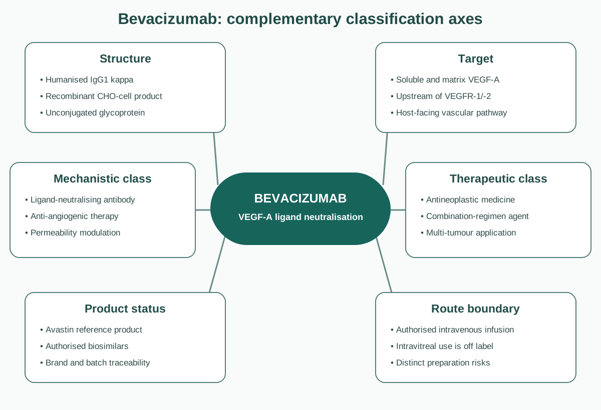
Figure 1. Bevacizumab is simultaneously a humanised IgG1, a VEGF-A ligand-neutralising agent, an anti-angiogenic oncology medicine and a biological reference product. Route and product context are separate axes: authorised intravenous Avastin and off-label intravitreal repackaging are not equivalent exposure systems.
Structural and engineering classification
Bevacizumab is a recombinant humanised IgG1 monoclonal antibody produced in Chinese hamster ovary cells. It was derived by humanising the murine anti-VEGF antibody A.4.6.1. Humanisation retained antigen-recognising residues while replacing most non-human sequence, reducing immunogenic potential without eliminating it.
As a glycoprotein, bevacizumab is defined by primary sequence, higher-order structure, glycosylation, charge distribution, aggregation profile, VEGF-binding activity and other quality attributes. An authorised biosimilar must demonstrate high similarity to the reference product through comparative analytical and functional evidence, supported by pharmacokinetic and clinical data as required. It is not evaluated as a conventional chemical generic.
Target and pathway classification
VEGF-A belongs to a broader VEGF family. Alternative splicing produces VEGF-A isoforms with different extracellular-matrix binding and distribution. Bevacizumab binds biologically active VEGF-A and prevents interaction with VEGFR-1 and VEGFR-2. It does not directly neutralise every VEGF-family ligand and does not directly inhibit the receptor kinase.
VEGFR-2 is the major mediator of endothelial proliferation, migration, survival and vascular permeability. VEGFR-1 has complex signalling and ligand-sequestering functions and is also expressed by non-endothelial cells. Neuropilins and matrix binding further shape the pathway. “Blocking angiogenesis” is therefore a systems-level consequence, not a simple on/off switch.
Physiological and tumour angiogenesis
Growing tumours can outstrip oxygen delivery. Hypoxia stabilises hypoxia-inducible factors, increasing transcription of VEGF and other adaptive genes. VEGF-A gradients stimulate endothelial activation, migration and proliferation, supporting new vascular sprouts. Tumour vessels are often disorganised, permeable and inefficient, producing heterogeneous perfusion and high interstitial pressure.
VEGF inhibition can suppress new-vessel formation, regress susceptible immature vessels and transiently “normalise” aspects of abnormal vasculature. Vascular normalisation may improve perfusion and delivery of concomitant therapy during a context-dependent window. Excessive or sustained pruning can also increase hypoxia and select adaptive tumour phenotypes. The effect cannot be reduced to “starving the tumour.”
Discovery and development history
The idea that tumour growth depends on angiogenesis was advanced by Judah Folkman and others in the 1970s. Subsequent isolation and cloning of vascular permeability factor/VEGF connected vascular leakage and endothelial proliferation to a definable molecular ligand. Napoleone Ferrara and colleagues developed neutralising anti-VEGF antibodies and showed inhibition of tumour growth in experimental models.
The murine antibody A.4.6.1 was humanised to create bevacizumab. Early clinical development explored whether a systemic antibody against a host-facing angiogenic ligand could be combined with cytotoxic chemotherapy. Unlike a tumour-antigen antibody, bevacizumab did not require expression of a single surface marker on malignant cells; the therapeutic hypothesis concerned the vascular support system shared by many solid tumours.
Pivotal colorectal-cancer evidence
The pivotal trial reported by Hurwitz and colleagues in 2004 compared irinotecan, bolus fluorouracil and leucovorin with or without bevacizumab in previously untreated metastatic colorectal cancer. Adding bevacizumab improved overall survival, progression-free survival and response. Hypertension and gastrointestinal perforation emerged as clinically important risks.
The US FDA approved bevacizumab in 2004 for use with chemotherapy in metastatic colorectal cancer. The European Union granted Avastin marketing authorisation on 12 January 2005. EU development subsequently expanded to defined settings in metastatic colorectal cancer, metastatic breast cancer, non-squamous non-small-cell lung cancer, advanced renal-cell cancer, epithelial ovarian/fallopian-tube/primary peritoneal cancer and persistent, recurrent or metastatic cervical cancer.
Evolution of the benefit–risk profile
Development across tumour types showed that anti-angiogenic benefit is regimen- and disease-specific. An efficacy result in one cancer cannot be extrapolated merely because VEGF is present in another. Regulatory decisions have differed by jurisdiction and indication as evidence evolved; notably, US accelerated approval for metastatic breast cancer was later withdrawn after reassessment, while defined breast-cancer indications remain authorised in the EU. This divergence illustrates that “approved” is not an intrinsic property of a molecule but a jurisdiction-, indication-, regimen- and time-specific conclusion.
Biosimilar era
Multiple bevacizumab biosimilars have been authorised in the EU. The totality-of-evidence approach supports authorised indications without repeating every reference-product outcome trial. Pharmacovigilance nevertheless requires product-level identification. Market share, indication mix, procurement switching and reporting stimulation can create apparent brand differences that are not biological differences.
The intravitreal boundary
Bevacizumab has been widely used off label by intravitreal injection for retinal vascular diseases because VEGF drives pathological ocular neovascularisation and permeability. Avastin is not formulated or authorised by EMA for intravitreal use. Small aliquots may be repackaged from oncology vials by pharmacies or compounding services.
This creates a distinct safety system: aseptic preparation, aliquot storage, syringe materials, transport, clustered endophthalmitis, silicone-oil droplets, ocular inflammation and product-source traceability become central. Systemic oncology data cannot simply be applied to the much smaller ocular dose, and ocular safety data cannot be used to dismiss systemic risks of intravenous oncology treatment.
Detailed mechanism of action
Bevacizumab binds VEGF-A and forms complexes that reduce free ligand available to activate VEGFR-1 and VEGFR-2. This is a pharmacological sink rather than irreversible elimination of VEGF production. Tumour cells, stromal cells and host tissues can continue to produce ligand, and pathway activity can recover as exposure declines or compensatory biology develops.
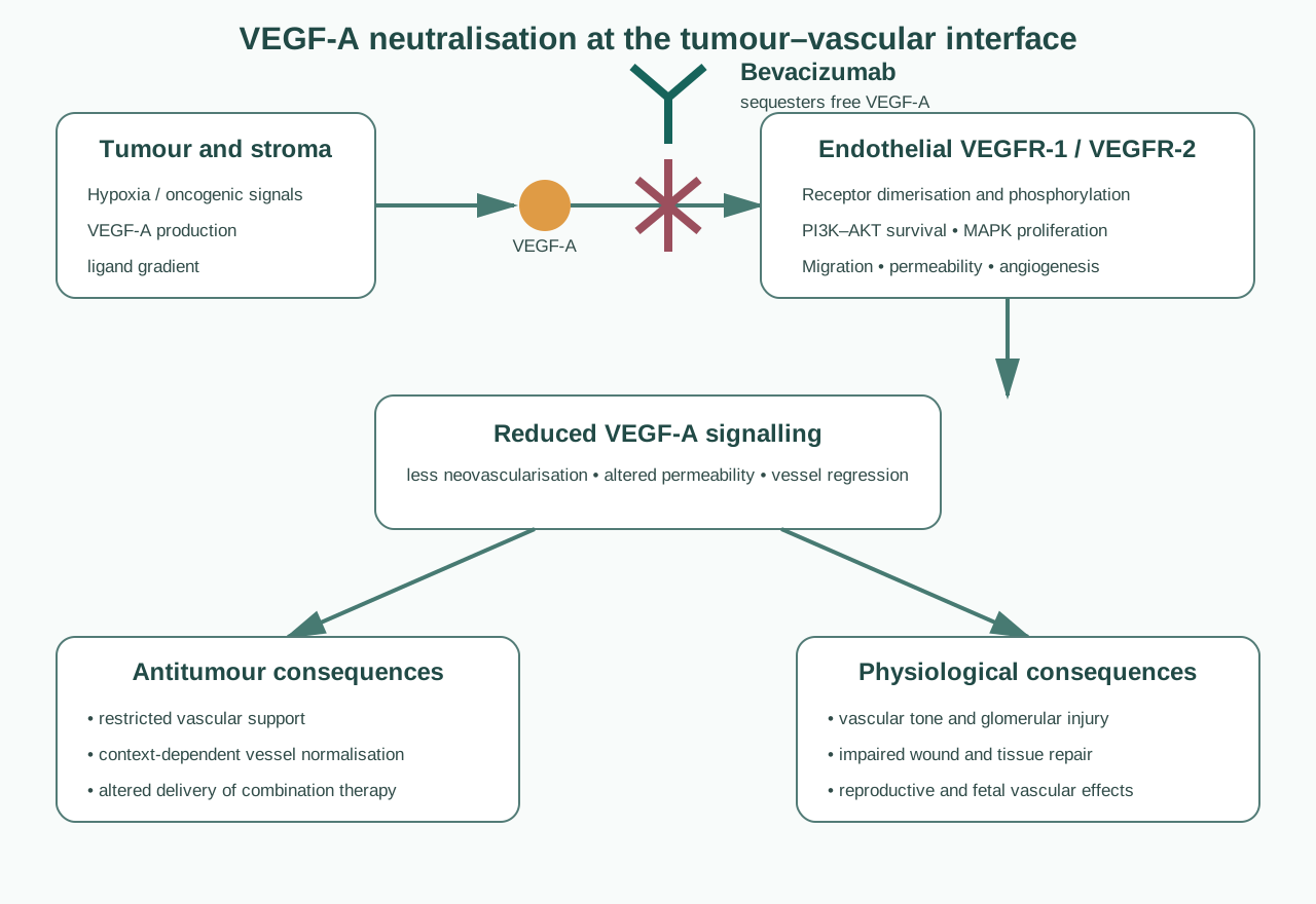
Figure 2. Hypoxic tumour and stromal cells release VEGF-A, which activates VEGFRs on endothelial cells to support angiogenesis, permeability and survival. Bevacizumab intercepts VEGF-A before receptor binding. Physiological use of the same pathway explains mechanism-linked vascular, renal, wound-healing and reproductive risks.
Ligand sequestration
Each bevacizumab molecule can engage VEGF-A through its antigen-binding arms. By lowering free VEGF-A, the antibody reduces receptor dimerisation and phosphorylation. Downstream effects include reduced signalling through pathways such as PLCγ–PKC–MAPK and PI3K–AKT, with less endothelial proliferation, migration, survival and permeability.
Bevacizumab does not directly kill tumour cells that lack a relevant dependence on VEGF autocrine signalling. Its principal established action is modification of the tumour–host vascular interface. Concomitant chemotherapy, immunotherapy or other targeted therapy supplies additional antitumour mechanisms.
Vascular regression and inhibition of neovascularisation
Immature tumour vessels can depend strongly on VEGF survival signals. Neutralisation may cause regression of susceptible vessels and inhibit formation of new ones. More mature vessels supported by pericytes and alternative signals may be less sensitive. Response therefore varies within and between tumours.
Vascular normalisation
Tumour vessels are abnormally leaky and poorly organised. Partial VEGF inhibition can reduce permeability, lower interstitial pressure and improve vascular architecture. Under some conditions this may enhance oxygenation or delivery of co-administered agents. The normalisation hypothesis is time- and dose-dependent and should not be presented as a universal clinical mechanism proven for every regimen.
Effects on vascular permeability
VEGF was initially described as vascular permeability factor. Reduced signalling tightens aspects of the endothelial barrier and can reduce oedema. This is relevant to tumour physiology and to the rapid radiographic changes sometimes observed in highly oedematous lesions. Reduced contrast enhancement may partly reflect altered permeability rather than equivalent reduction in viable tumour—a potential imaging-response complication.
Host-vascular consequences
Physiological VEGF maintains endothelial nitric-oxide signalling and microvascular homeostasis. Its inhibition can increase vascular resistance and blood pressure. In the kidney, podocyte-derived VEGF supports glomerular endothelium; disruption can produce proteinuria and thrombotic-microangiopathy-like injury. During tissue repair, angiogenesis supports granulation and wound healing. In reproductive tissues and the developing fetus, VEGF signalling is essential for vascular development and organ function.
These effects connect molecular mechanism to the safety profile more directly than a list of adverse-reaction frequencies.
Adaptation and resistance
Tumours may evade VEGF-A neutralisation by increasing ligand production, recruiting alternative pro-angiogenic pathways, co-opting existing vessels, adopting invasive growth, increasing pericyte support or changing myeloid and stromal populations. Host and tumour mechanisms interact. No validated universal biomarker reliably selects all patients who will benefit from bevacizumab across indications.
Pharmacokinetics and exposure
Bevacizumab has a long half-life typical of an IgG antibody and is administered intravenously at weight-based, indication-specific doses and intervals. Clearance varies with body size, sex, albumin, tumour burden and other factors. The long persistence means that vascular and wound-healing considerations continue after the final dose.
Pharmacokinetic interpretation differs radically for intravitreal exposure. Ocular administration produces local concentrations from a small dose but some systemic absorption occurs. Route, dose, formulation handling and patient population must be recorded before comparing events.
Major safety risks
Gastrointestinal perforation and fistulae
Gastrointestinal perforation is among the most serious bevacizumab risks and can be fatal. Contributing biology may include impaired microvascular repair, tumour necrosis involving bowel, ischaemia and delayed healing. Risk is shaped by tumour site, bowel involvement, inflammation, obstruction, prior surgery, radiation and concomitant medicines.
Fistulae can involve gastrointestinal, genitourinary, respiratory or other structures. Cervical-cancer populations previously exposed to pelvic radiation require particular contextual assessment. Reports should document anatomical site, tumour involvement, imaging, surgery/radiation history, infection, intervention and outcome.
Wound-healing complications
Angiogenesis is essential to wound repair. Bevacizumab can cause dehiscence, delayed healing, anastomotic complications, abscess and other postoperative morbidity. Current product information specifies timing precautions around major surgery and resumption only after adequate healing.
PV assessment requires exact dates of the last dose, procedure, complication and any restart; procedure type and urgency; wound status before treatment; infection; nutrition; steroids; diabetes; radiation and surgical management. “Postoperative complication” without this chronology is not evaluable.
Haemorrhage
Bleeding ranges from minor mucosal events such as epistaxis to serious or fatal pulmonary, gastrointestinal, central-nervous-system or tumour-associated haemorrhage. In non-small-cell lung cancer, predominantly squamous histology and central/cavitating tumours informed efforts to reduce severe pulmonary-haemorrhage risk.
Case review should capture site, grade, imaging/endoscopy, tumour erosion, platelet count, coagulation, anticoagulants or antiplatelets, recent procedures and radiation. Anti-VEGF exposure may be contributory even when local tumour anatomy is the immediate source.
Arterial and venous thromboembolism
Arterial events include stroke, transient ischaemic attack, myocardial infarction and other arterial thromboses. Older age, diabetes and prior arterial thromboembolism can increase risk. Venous thromboembolism is also reported in already high-risk cancer populations.
Separate arterial from venous events in aggregate analysis. Record cancer activity, immobility, central lines, surgery, thrombophilia, cardiovascular history and concomitant treatment. A crude “thrombosis” grouping loses clinically meaningful mechanism and risk-factor information.
Hypertension
Hypertension is common and mechanistically consistent with reduced nitric-oxide signalling and microvascular rarefaction. It can be severe and may contribute to cardiac, renal or neurological complications. Baseline blood pressure, serial values, antihypertensive treatment, adherence and evidence of end-organ injury are more useful than a narrative diagnosis alone.
Proteinuria and renal injury
Proteinuria ranges from low-grade findings to nephrotic syndrome. Renal biopsy in severe cases may show thrombotic microangiopathy. Monitoring and interruption criteria are defined in current product information. Cases should include baseline renal disease, serial quantitative protein assessment, creatinine, blood pressure, oedema, biopsy if performed and competing nephrotoxic therapy.
Posterior reversible encephalopathy syndrome
PRES is rare and presents with combinations of seizure, headache, altered mental status, visual disturbance and hypertension. Magnetic-resonance imaging commonly demonstrates vasogenic oedema, often posteriorly, but patterns vary and “reversible” is not guaranteed. Differential diagnoses include brain metastasis, stroke, infection, metabolic disturbance and chemotherapy toxicity.
Congestive heart failure
Cardiac failure has been observed, particularly in patients previously exposed to anthracyclines or chest-wall radiation and in some breast-cancer regimens. Bevacizumab may add vascular load through hypertension or endothelial effects. Full cardiac and treatment history is required rather than attributing the event from chronology alone.
Ovarian failure, pregnancy and fetal risk
VEGF is important in ovarian angiogenesis. Ovarian failure has been observed in premenopausal patients and may not always reverse. Menstrual history, ovarian reserve measures where available, fertility counselling, chemotherapy and recovery should be documented.
Bevacizumab is contraindicated in pregnancy in EU product information. VEGF inhibition can disrupt fetal vascular development, and an IgG antibody can cross the placenta. Contraception and post-treatment intervals must follow current product information. Pregnancy follow-up requires dose dates, gestational timing, co-therapy, ultrasound findings, outcome and infant assessment.
Infusion reactions and hypersensitivity
Reactions can include dyspnoea, flushing, rash, blood-pressure changes, chest discomfort and hypersensitivity manifestations. Exact timing, infusion number and rate, treatment, response to interruption and alternative causes are necessary. Anti-drug antibodies are uncommon but assay and clinical context matter.
Pharmacovigilance architecture
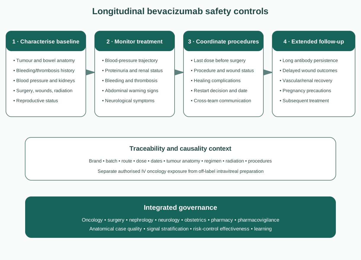
Figure 3. Bevacizumab safety management links baseline anatomy and vascular risk, treatment monitoring, surgery/wound coordination and extended follow-up. Exact product, batch, route, regimen and timing support every stage.
The central challenge is to preserve anatomy and chronology. Perforation, bleeding and fistulae cannot be understood without tumour location, radiation and surgery. Hypertension, proteinuria and PRES require serial measurements. Wound events require the interval between dose and procedure. Pregnancy risk requires gestational timing and residual exposure.
| Domain | High-value information |
|---|---|
| Product | Brand, batch, concentration, route, dose, dates and country |
| Cancer | Primary site, histology, stage, tumour invasion, bowel or vessel involvement |
| Regimen | All antineoplastic medicines, radiation, anticoagulants and corticosteroids |
| Procedures | Operation type, wound status and exact perioperative exposure intervals |
| Vascular baseline | Blood pressure, renal function/proteinuria, thrombosis and cardiovascular history |
| Event | Anatomy, imaging/pathology, serial laboratory values, intervention and outcome |
| Reproductive | Menstrual/fertility baseline, contraception, conception and gestational exposure |
Event-specific assessment
For perforation or fistula, reconstruct tumour anatomy, inflammatory or obstructive disease, radiation, endoscopy and surgery. For haemorrhage, identify the source and local lesion as well as systemic haemostatic factors. For thrombosis, distinguish arterial and venous events and document conventional cancer-associated risks.
For hypertension and proteinuria, serial values establish onset, severity, response and recurrence. A single coded term cannot reveal whether PRES, thrombotic microangiopathy or cardiac failure formed part of the same vascular syndrome. For wound complications, dose-to-surgery and surgery-to-restart intervals are indispensable.
Off-label intravitreal cases
Ocular reports require laterality, indication, injected dose/volume, vial and aliquot source, compounder, syringe, preparation date, storage, injection date, other patients exposed to the same lot and microbiological results. Clustering is a critical clue to contamination. Systemic vascular events after intravitreal use require cautious assessment because baseline vascular disease is common and systemic exposure differs from oncology dosing.
Biosimilars and traceability
Authorised bevacizumab biosimilars are expected to have no clinically meaningful differences from Avastin in their authorised uses. Nevertheless, brand and batch capture are required for biological traceability and quality investigation. “Bevacizumab” alone may prevent analysis of a batch-linked cluster or switching sequence.
Spontaneous counts cannot establish comparative incidence. Uptake, market share, indication mix, stimulated reporting, hospital procurement and missing brand data must be considered. Delayed events should be mapped across all products received rather than automatically assigned to the latest brand.
Risk minimisation and effectiveness
Before treatment, clinicians should evaluate tumour anatomy, bleeding and thrombotic history, blood pressure, renal status, recent or planned surgery, wound healing and reproductive status according to current product information. During treatment, blood pressure, urine protein, clinical bleeding, abdominal symptoms, neurological symptoms and wound status require appropriate surveillance.
Treatment planning must coordinate oncology and surgical teams. A medicine reconciliation that omits bevacizumab because it is an “infusion” can lead to unsafe procedural timing. Patients need actionable education about severe abdominal pain, bleeding, neurological symptoms and wound problems.
Effectiveness measures can include completeness of baseline blood pressure and proteinuria assessment; adherence to perioperative intervals; timeliness of evaluation for abdominal or neurological symptoms; product/batch capture; contraception documentation; and evidence that serious events produce system learning. Distribution of written warnings alone does not demonstrate effectiveness.
Inspection and governance perspective
An inspectable system should show:
- current product-specific safety information and indication/regimen control;
- brand, batch and route retention, including biosimilar switches;
- event-specific follow-up for perforation, fistula, bleeding, thrombosis, hypertension, proteinuria, PRES, wound healing and pregnancy;
- exact surgery–exposure chronology;
- stratification by tumour type, anatomy, regimen and radiation;
- integration of serial blood-pressure, renal and neurological evidence;
- separation of authorised intravenous use from off-label intravitreal preparation;
- quality-defect escalation for clustered ocular or infusion events;
- exposure-aware interpretation of brand reporting patterns;
- traceability from signal evaluation to risk-management action.
Common failures include coding “abdominal pain” without determining perforation; combining arterial and venous thromboses; omitting tumour invasion at the bleeding site; accepting “recent surgery” without dates; reporting proteinuria without quantification; assuming PRES is always reversible; and treating intravitreal compounded aliquots as if their preparation chain were identical to an intact oncology vial.
Practical checklist
- Confirm exact indication, regimen, product, batch, dose and route.
- Record tumour site, histology, bowel/vessel involvement and prior radiation.
- Capture baseline and serial blood pressure, urine protein and renal function.
- Reconstruct all operations and wounds against bevacizumab dose dates.
- Phenotype bleeding, thrombosis, perforation and fistula anatomically.
- Investigate headache, seizure, visual change or confusion urgently for PRES and alternatives.
- Document fertility counselling, pregnancy prevention and gestational exposure.
- For intravitreal reports, capture the full aliquoting and injection chain.
- Analyse biosimilar reports with exposure and reporting biases, not counts alone.
- Maintain surveillance after discontinuation because exposure and repair effects persist.
Key takeaways
Bevacizumab is a humanised IgG1 monoclonal antibody that neutralises VEGF-A upstream of VEGFR-1 and VEGFR-2. It modifies the tumour–host vascular interface rather than requiring direct binding to a tumour-cell antigen.
Its mechanism includes ligand sequestration, inhibition of neovascularisation, regression of susceptible immature vessels, reduced permeability and context-dependent vascular normalisation. Tumours can adapt through alternative angiogenic and vessel-use strategies.
Physiological VEGF explains the safety pattern: hypertension and proteinuria reflect vascular and glomerular effects; perforation, fistulae and wound complications reflect impaired tissue repair; haemorrhage and thrombosis reflect disturbed vascular integrity; reproductive and fetal risks reflect developmental angiogenesis.
PV assessment depends on anatomy, serial measurements and exact chronology. Product, regimen, radiation, surgery and baseline vascular risk must be evaluated together.
Authorised intravenous oncology use and off-label intravitreal use are separate exposure systems. Biosimilars add a further need for brand and batch traceability without implying clinically meaningful differences.
References
- European Medicines Agency. Avastin: EPAR. Product information and assessment history.
- European Medicines Agency. Avastin: EPAR product information. Revision accessed September 2026.
- Ferrara N, Hillan KJ, Gerber HP, Novotny W. Discovery and development of bevacizumab, an anti-VEGF antibody for treating cancer. Nat Rev Drug Discov. 2004;3:391–400. doi:10.1038/nrd1381.
- Kim KJ, Li B, Winer J, et al. Inhibition of vascular endothelial growth factor-induced angiogenesis suppresses tumour growth in vivo. Nature. 1993;362:841–844. doi:10.1038/362841a0.
- Presta LG, Chen H, O'Connor SJ, et al. Humanization of an anti-vascular endothelial growth factor monoclonal antibody for the therapy of solid tumors and other disorders. Cancer Res. 1997;57(20):4593–4599.
- Hurwitz H, Fehrenbacher L, Novotny W, et al. Bevacizumab plus irinotecan, fluorouracil, and leucovorin for metastatic colorectal cancer. N Engl J Med. 2004;350:2335–2342. doi:10.1056/NEJMoa032691.
- Sandler A, Gray R, Perry MC, et al. Paclitaxel-carboplatin alone or with bevacizumab for non-small-cell lung cancer. N Engl J Med. 2006;355:2542–2550. doi:10.1056/NEJMoa061884.
- Miller K, Wang M, Gralow J, et al. Paclitaxel plus bevacizumab versus paclitaxel alone for metastatic breast cancer. N Engl J Med. 2007;357:2666–2676. doi:10.1056/NEJMoa072113.
- Tewari KS, Sill MW, Long HJ, et al. Improved survival with bevacizumab in advanced cervical cancer. N Engl J Med. 2014;370:734–743. doi:10.1056/NEJMoa1309748.
- Ferrara N, Adamis AP. Ten years of anti-vascular endothelial growth factor therapy. Nat Rev Drug Discov. 2016;15:385–403. doi:10.1038/nrd.2015.17.
- Jain RK. Normalization of tumor vasculature: an emerging concept in antiangiogenic therapy. Science. 2005;307:58–62. doi:10.1126/science.1104819.
- Carmeliet P, Jain RK. Molecular mechanisms and clinical applications of angiogenesis. Nature. 2011;473:298–307. doi:10.1038/nature10144.
- Eremina V, Jefferson JA, Kowalewska J, et al. VEGF inhibition and renal thrombotic microangiopathy. N Engl J Med. 2008;358:1129–1136. doi:10.1056/NEJMoa0707330.
- Ranpura V, Hapani S, Wu S. Treatment-related mortality with bevacizumab in cancer patients: a meta-analysis. JAMA. 2011;305:487–494. doi:10.1001/jama.2011.51.
- Hapani S, Chu D, Wu S. Risk of gastrointestinal perforation in patients with cancer treated with bevacizumab: a meta-analysis. Lancet Oncol. 2009;10:559–568. doi:10.1016/S1470-2045(09)70112-3.
- Scappaticci FA, Fehrenbacher L, Cartwright T, et al. Surgical wound healing complications in metastatic colorectal cancer patients treated with bevacizumab. J Surg Oncol. 2005;91:173–180. doi:10.1002/jso.20301.
- European Medicines Agency. Guideline on similar biological medicinal products. CHMP/437/04 Rev 1.
- European Medicines Agency. Avzivi: EPAR. Biosimilar assessment overview.
Regulatory Note
This article is an educational scientific and pharmacovigilance review, not prescribing advice. Authorised indications, regimens, contraindications, monitoring, perioperative intervals, pregnancy precautions and regulatory status vary by product and jurisdiction and may change. Consult current product-specific information. Avastin is authorised for intravenous oncology use; intravitreal administration is off label in the EU and may involve repackaging or compounding with separate quality, traceability and clinical-governance requirements.