Blinatumomab: CD19–CD3 T-cell engagement, acute lymphoblastic leukaemia, and product pharmacovigilance
- Blinatumomab: CD19–CD3 T-cell engagement, acute lymphoblastic leukaemia, and product pharmacovigilance
- Why this molecule changed the idea of an antibody
- Classification: what blinatumomab is—and is not
- First principles: how antibodies recognize a target
- The two targets
- From bridge to cytolytic synapse
- B-cell precursor acute lymphoblastic leukaemia from first principles
- Remission is not always eradication
- How the mechanism changes across authorised indications
- Treatment map: other ways to attack the disease
- The wider CD3-engager class
- Why continuous infusion is necessary
- Mechanism-linked safety
- Product Pharmacovigilance
- Evolution of the safety system
- What the medicine solves—and what it does not
- Official regulatory reference material
- References
- Regulatory Note
Why this molecule changed the idea of an antibody
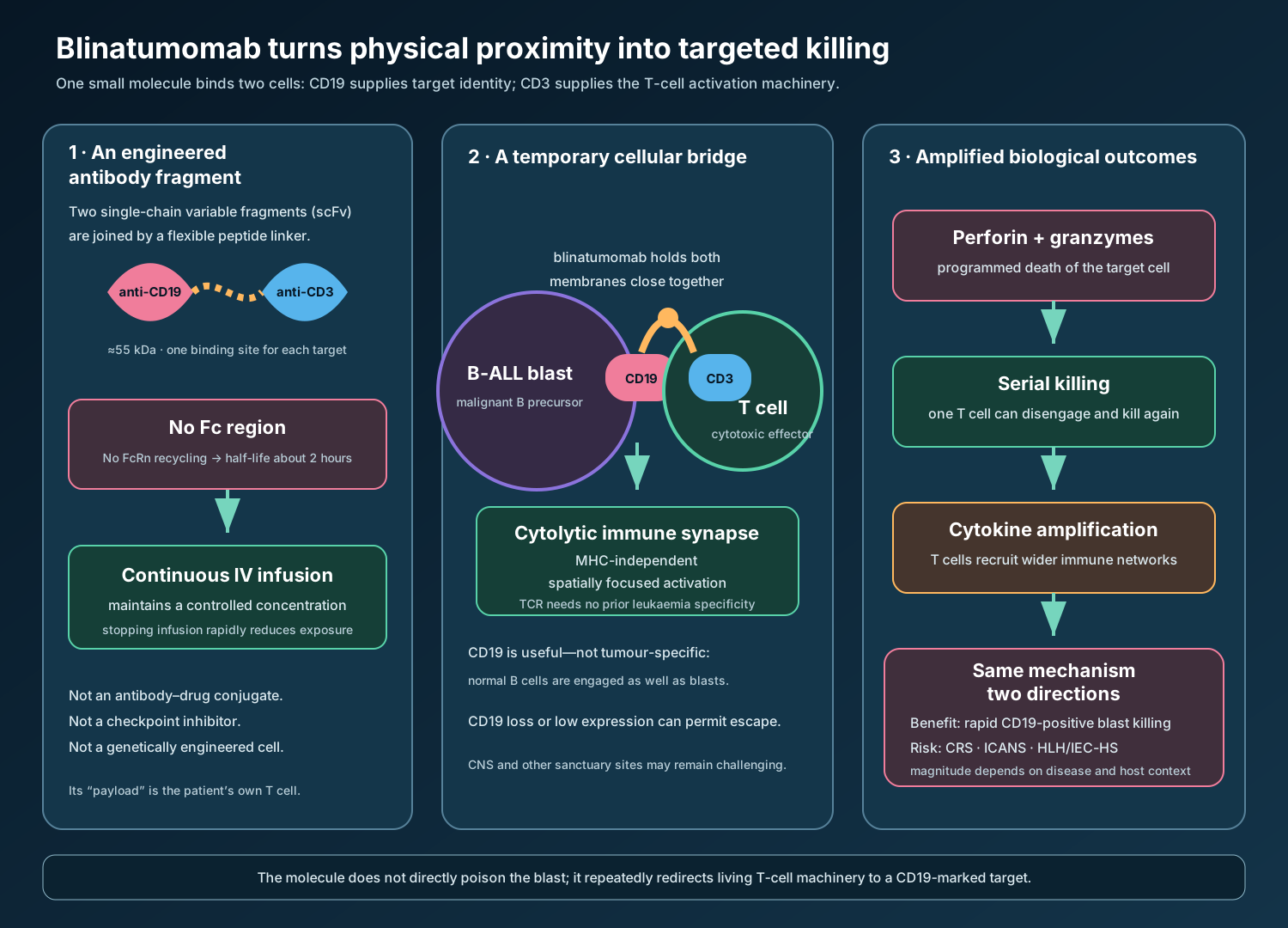
Blinatumomab does not behave like a conventional monoclonal antibody. A typical therapeutic immunoglobulin G (IgG) recognizes one target and retains a long-lived Fc region. Blinatumomab is a much smaller, fragment-based bispecific T-cell engager. One binding end recognizes CD19 on B-lineage cells; the other recognizes CD3 on T lymphocytes. It acts as a temporary molecular bridge, bringing a patient’s own cytotoxic T cell into direct contact with a leukaemia cell.[1–4]
That bridge can be highly effective, but it is deliberately incomplete. Blinatumomab lacks the Fc region that normally gives IgG antibodies much of their stability and long circulation time. Its elimination half-life is only about two hours, so therapeutic exposure requires continuous intravenous infusion. The same close, sustained T-cell activation that kills malignant cells can also produce cytokine release syndrome (CRS), neurological toxicity including immune effector cell-associated neurotoxicity syndrome (ICANS), and—in uncommon cases—an HLH-like hyperinflammatory state.
The medicine therefore cannot be understood by memorizing a list of warnings. Its structure explains its infusion method; its mechanism explains both benefit and harm; and its narrow microgram-dose delivery system explains why preparation, pump, tubing, and line-flushing errors are pharmacovigilance concerns.
Classification: what blinatumomab is—and is not
| Classification axis | Blinatumomab | Why the distinction matters |
|---|---|---|
| Molecular structure | Two single-chain variable fragments joined by a flexible linker | Much smaller than intact IgG and lacks an Fc region |
| Binding design | Bispecific: CD19 × CD3 | Connects two different cells rather than blocking one soluble mediator |
| Functional class | T-cell engager; historically termed a BiTE construct | Redirects endogenous T cells to a chosen surface antigen |
| Therapeutic class | Antineoplastic immunotherapy; ATC L01FX07 in the EU | Its characteristic toxicities arise from immune activation, not only cytotoxic chemotherapy |
| Targeted lineage | CD19-positive B-lineage cells | Normal B cells are depleted alongside malignant B-cell precursors |
| Administration | Continuous intravenous infusion in repeated cycles | Short half-life makes delivery continuity part of pharmacology and safety |
It is not an antibody–drug conjugate: it carries no cytotoxic payload. It is not a checkpoint inhibitor: it does not release an inhibitory brake such as PD-1. It is not CAR-T therapy: no patient cells are removed, genetically engineered, expanded, and reinfused. It is also not a conventional full-length bispecific IgG. These platforms may all recruit immunity against cancer, but their persistence, manufacturing, target geometry, and risk-control systems differ.
First principles: how antibodies recognize a target
A conventional antibody resembles a Y. Two heavy chains and two light chains create paired antigen-binding tips, while the Fc stem interacts with immune receptors and the neonatal Fc receptor, which recycles IgG and prolongs its circulation.
Each binding tip contains variable domains from one heavy chain and one light chain. Molecular engineers can connect those two variable domains with a short peptide linker to form a single-chain variable fragment (scFv). Blinatumomab joins an anti-CD19 scFv to an anti-CD3 scFv. The result is a single polypeptide of roughly 55 kDa—about one-third the mass of an intact IgG—with one functional binding site for each target.[3,4]
Removing Fc produces several consequences:
- killing depends mainly on recruited T cells, not Fc-receptor-bearing natural killer cells or complement;
- there is no FcRn-mediated recycling to sustain the usual multi-week IgG half-life;
- drug concentration can fall quickly when infusion stops, which helps make some toxicities reversible but creates vulnerability to interruptions;
- continuous delivery and exact microgram-level dosing become integral to the product’s effect.
The two targets
CD19: a B-lineage identity marker
CD19 is a transmembrane co-receptor expressed from early B-cell development through most mature B cells. It amplifies signals from the B-cell receptor, allowing a B cell to respond efficiently to antigen. Haematopoietic stem cells and most plasma cells do not express CD19, but B-cell precursor acute lymphoblastic leukaemia (BCP-ALL) blasts usually do.
This distribution is therapeutically useful: CD19 marks the malignant lineage while sparing stem cells that can rebuild blood formation. It is not tumour-specific. Normal CD19-positive B cells are also removed, causing B-cell aplasia and reduced immunoglobulin concentrations. Loss or reduction of CD19 on surviving leukaemia cells can allow immune escape.
CD3: the T-cell signalling gateway
CD3 is a multi-chain signalling complex associated with the T-cell receptor (TCR). The TCR recognizes a peptide presented by a major histocompatibility complex molecule; CD3 carries that recognition signal across the cell membrane. Blinatumomab binds the CD3 epsilon component and brings the signalling machinery close to a CD19-positive cell.
This recruitment is MHC-independent and does not require the TCR to have been naturally selected for a leukaemia antigen. The antibody supplies physical specificity: “activate here, beside this CD19-positive cell.” It does not make T-cell biology indiscriminate; activation is strongest when both ends engage and a productive cell–cell interface forms.
From bridge to cytolytic synapse
The contact zone between a cytotoxic T cell and its target is an immune synapse—an organized interface that focuses killing signals. Blinatumomab stabilizes proximity between CD3 and CD19. T cells then reorganize their cytoskeleton and release granules containing perforin and granzymes. Perforin helps deliver granzymes into the target; granzymes initiate programmed cell death.
The activated T cell also proliferates and releases cytokines. After one target dies, it can disengage and attack another: serial killing. This amplification explains why very low blinatumomab concentrations can have substantial biological effects. It also explains why the amount of leukaemia, the number and fitness of available T cells, CD19 density, inhibitory signals in the microenvironment, and anatomical sanctuary sites all influence response.
B-cell precursor acute lymphoblastic leukaemia from first principles
Blood cells arise from haematopoietic stem and progenitor cells in bone marrow. Normal B-cell development passes through controlled precursor stages. In BCP-ALL, acquired genetic changes arrest differentiation and promote survival and proliferation. Immature blasts accumulate instead of maturing into functional B cells.
The marrow is a finite production space. As blasts expand, they displace normal precursors:
- reduced red-cell production causes anaemia, fatigue, and breathlessness;
- reduced functional neutrophils increases infection risk;
- reduced platelets cause bruising and bleeding;
- blasts can enter blood and infiltrate lymph nodes, liver, spleen, testes, or the central nervous system (CNS).
The Philadelphia chromosome is a translocation that creates the BCR::ABL1 fusion kinase. It supplies a continuously active growth signal and defines a biologically important subgroup. Tyrosine-kinase inhibitors (TKIs) directly suppress BCR::ABL1, whereas blinatumomab acts at the surface CD19 marker. These mechanisms can be complementary, but authorisations specify different settings and prior-treatment requirements.
Remission is not always eradication
Morphological complete remission generally means that marrow blasts are below the threshold visible by routine microscopy, blood counts recover, and there is no obvious extramedullary disease. A marrow containing fewer than 5% blasts can still harbour millions of leukaemia cells.
Measurable residual disease (MRD) uses flow cytometry, fusion-transcript testing, immunoglobulin/TCR rearrangements, or next-generation sequencing to detect disease at much greater sensitivity—often one abnormal cell among 10,000 to one million cells, depending on the validated assay. Persistent MRD is a strong relapse marker.
This creates an ideal biological setting for a T-cell engager: the target population is small enough to reduce explosive immune activation, yet clinically important enough to justify eradication. MRD negativity is not synonymous with cure, because sampling and assay sensitivity have limits and sanctuary sites may remain.
How the mechanism changes across authorised indications
Relapsed or refractory CD19-positive BCP-ALL
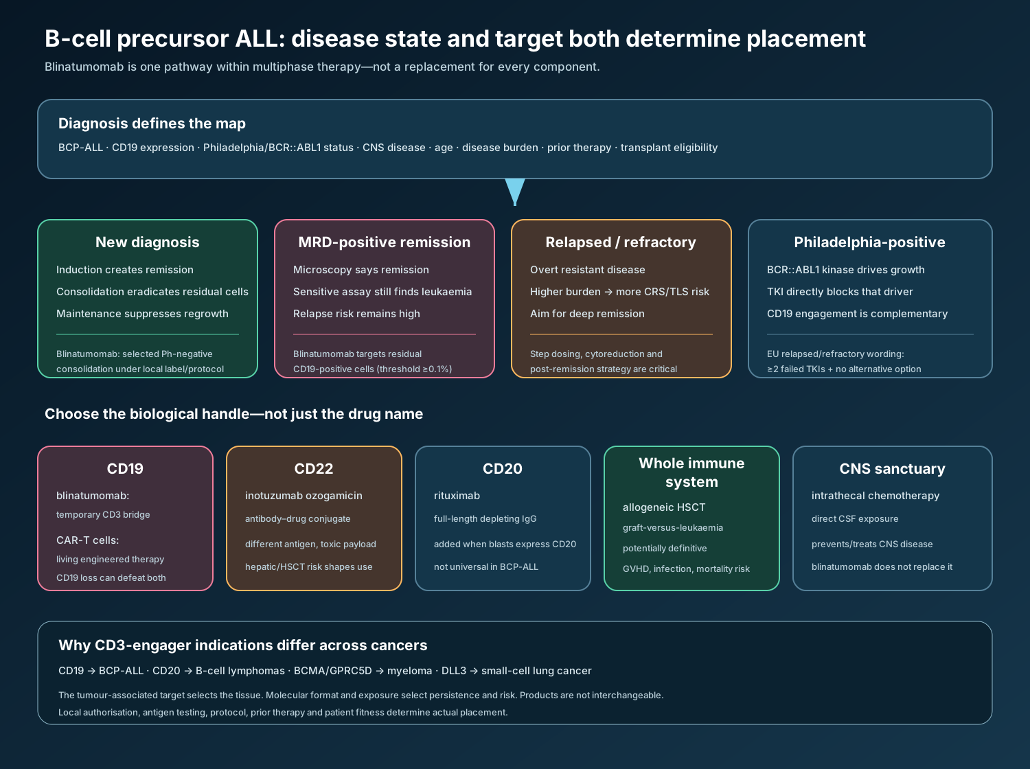
Relapsed disease returns after remission; refractory disease fails to enter remission with prior therapy. Both settings often contain a high blast burden, resistant subclones, injured marrow reserve, and T cells altered by previous therapy. The immediate objective is usually to induce another deep remission, often as a bridge to allogeneic haematopoietic stem-cell transplantation (HSCT) or another definitive strategy.
The phase 3 TOWER trial randomized 405 adults with Philadelphia-negative relapsed or refractory BCP-ALL to blinatumomab or investigator-selected chemotherapy. Median overall survival was 7.7 versus 4.0 months (hazard ratio 0.71); complete remission with full or partial haematological recovery was 42% versus 20%.[5,6] The survival curve later converged, illustrating that response is not equivalent to durable cure and that post-remission strategy matters.
In Philadelphia-positive relapsed or refractory disease, the single-arm ALCANTARA study supported activity after TKI failure. US labelling includes relapsed or refractory CD19-positive BCP-ALL without a Philadelphia-specific prior-TKI qualifier. EU labelling specifies that adults with Philadelphia-positive disease should have failed at least two TKIs and have no alternative treatment option.[1,2]
High disease burden increases both the number of target encounters and cytokine release. Cytoreduction, dexamethasone premedication, inpatient initiation, and step dosing during the first relapsed/refractory cycle are therefore not administrative details; they are mechanism-based risk controls.
MRD-positive first or second remission
Here the patient appears to be in complete remission by morphology but has MRD of at least 0.1%, the labelled threshold. The BLAST study found complete MRD response during the first cycle in 78% of evaluable adults.[7,8] Responders had better survival than non-responders, but that comparison was not randomized and cannot prove that clearing MRD alone caused the survival difference.
The therapeutic purpose is to deepen remission and reduce relapse risk, potentially before HSCT. Lower target burden generally reduces CRS frequency compared with overt relapsed disease, but it does not abolish neurological, infectious, or infusion-related risk.
Newly diagnosed Philadelphia-negative disease in consolidation
Induction chemotherapy aims to produce remission. Consolidation treats disease that is no longer morphologically visible but could regrow. Maintenance then suppresses residual clones over a longer period. Blinatumomab cycles can be intercalated with chemotherapy during consolidation.
E1910 changed the drug’s place in adult care. Among 224 adults aged 30–70 years who were MRD-negative after induction/intensification, adding blinatumomab to consolidation improved three-year overall survival from about 68–69% to about 85% and relapse-free survival from 64% to 80%.[9,10] Benefit in already MRD-negative patients demonstrates that a negative assay is a threshold statement, not proof that every leukaemia cell has gone.
The 2024 US consolidation approval also used a paediatric/young-adult high-risk first-relapse study: five-year overall survival was 78.4% with blinatumomab consolidation versus 41.4% with intensive chemotherapy, and five-year relapse-free survival was 61.1% versus 27.6%.[10] The EU indication is narrower by population wording: adult newly diagnosed Philadelphia-negative CD19-positive BCP-ALL consolidation, and paediatric high-risk first-relapse consolidation.[1]
Paediatric disease
Children are not small adults. Weight- or body-surface-area dosing, developing organs, different genetic subtypes, treatment sequencing, and late effects all matter. Both regions include children from one month of age in specified settings, but the exact indication language differs.[1,2]
The EU RMP treats developmental impairment, later CNS relapse, long-term toxicity, secondary malignancy, and HSCT-related toxicity in children as potential or missing-information questions requiring long-term observation.[3] These categories do not assert that blinatumomab causes each outcome. They identify evidence gaps important enough for structured follow-up.
Treatment map: other ways to attack the disease
| Strategy | Biological route | Where it contributes | Central limitation |
|---|---|---|---|
| Multi-agent chemotherapy | Damages DNA, mitosis, nucleotide synthesis, or cell survival | Induction, consolidation and maintenance backbones | Non-selective tissue toxicity and resistant clones |
| BCR::ABL1 TKI | Blocks the Philadelphia fusion kinase | Philadelphia-positive ALL | Requires the driver; resistance mutations and residual disease remain possible |
| Blinatumomab | Bridges CD19 to CD3 | MRD, consolidation, and relapsed/refractory settings per local label | Continuous infusion; CRS, ICANS, infection; CD19 escape |
| Inotuzumab ozogamicin | CD22-directed antibody–drug conjugate delivers calicheamicin | Relapsed/refractory BCP-ALL | Hepatic sinusoidal obstruction risk, especially around HSCT |
| CD19 CAR-T cells | Genetically programmed living T-cell therapy | Selected relapsed/refractory patients | Manufacturing time, prolonged cellular persistence, CRS/ICANS, access |
| Rituximab | Full IgG against CD20 | Added to chemotherapy when leukaemia expresses CD20 | CD20 is not expressed on every BCP-ALL blast |
| Allogeneic HSCT | Replaces haematopoiesis and adds graft-versus-leukaemia immunity | Selected high-risk or relapsed patients in remission | Transplant mortality, graft-versus-host disease, infection |
| CNS-directed intrathecal therapy | Places chemotherapy into cerebrospinal fluid | Prevents or treats CNS sanctuary disease | Blinatumomab does not replace it |
Target choice follows antigen expression and disease biology. CD19, CD20, and CD22 are not interchangeable labels; their density and persistence differ across B-cell maturation and after therapy. A CD19-negative relapse may still express CD22. A BCR::ABL1-positive clone requires attention to kinase control even when it also expresses CD19.
The wider CD3-engager class
Blinatumomab was the first widely authorised bispecific T-cell engager, but CD3 recruitment is now used across malignancies. CD20×CD3 antibodies treat selected B-cell lymphomas; BCMA×CD3 and GPRC5D×CD3 antibodies treat multiple myeloma; DLL3×CD3 engagement is used in small-cell lung cancer. The second target determines which tissue is brought to the T cell, while molecular format and exposure determine how long and where engagement occurs.
Shared CD3 activation produces class-level overlap—CRS, neurological events, infection and cytopenias—but frequency and management cannot be copied mechanically between products. A long-lived full IgG bispecific given intermittently and a short-lived Fc-free construct infused continuously are pharmacologically different.
Why continuous infusion is necessary
With a half-life near two hours, intermittent injection would create a high peak followed by rapid loss of activity. Continuous infusion aims for a controlled steady concentration. A typical cycle contains 28 treatment days followed by a 14-day treatment-free interval; cycle number, dose, step-up schedule, hospital observation, and bag duration depend on indication, weight, cycle, jurisdiction, and current product information.[1,2]
Dexamethasone before initiation or restart reduces early immune activation. Intrathecal chemotherapy is required or recommended to prevent CNS relapse because systemic T-cell engagement is not a substitute for validated CNS-directed treatment. In high-burden relapsed/refractory disease, cytoreduction and anti-hyperuricaemic measures reduce tumour-lysis risk.
The infusion line is a pharmacological reservoir. It should use a dedicated lumen and must not be flushed at bag change or treatment completion: flushing can deliver the residual line content as an unintended bolus. Bags require exact preparation and programmed rates, and interruption length determines whether supervised restart and repeat premedication are needed. Home infusion transfers part of the safety system to pumps, caregivers, home-care nurses, pharmacies, and emergency communication.
Mechanism-linked safety
Cytokine release syndrome
Activated T cells release interferon-gamma, tumour-necrosis factor and other mediators; recruited monocytes and macrophages can amplify interleukin-6 and inflammatory networks. When activation becomes systemic, fever, hypotension, hypoxia, capillary leak, coagulopathy, and organ dysfunction may follow. This is CRS: an intended immune mechanism exceeding the safe physiological envelope.
Median onset is about two days. In the current US label, CRS was reported in 15% of relapsed/refractory patients, 7% of MRD-positive patients, and 16% during consolidation, using a broad group of overlapping terms.[2] The EU RMP’s larger pooled analysis reports 18.9% across 1,537 ALL study participants, with the greatest risk on day 2.[3] Different datasets and definitions explain why a single “incidence” should not be quoted without context.
CRS overlaps clinically with infusion reactions, infection, capillary leak, tumour lysis and HLH/IEC-HS. Blood cultures and antimicrobial decisions cannot wait merely because CRS is plausible. Severe CRS requires infusion interruption and corticosteroid-based management; life-threatening CRS requires permanent discontinuation under US labelling.[2]
Neurological toxicity and ICANS
Headache and tremor are common; severe manifestations include aphasia, dysgraphia, confusion, encephalopathy, impaired consciousness, ataxia, cranial-nerve abnormalities, and seizures. The US clinical-trial incidence of neurological toxicity is approximately 65%, with grade 3 or higher events around 13%; signs consistent with ICANS occurred in 7.5%.[2] Most events resolve after interruption, but fatal events and persistent harm have occurred.
The mechanism is incompletely defined. CD19 is not a neuronal target. Immune-cell adhesion to activated endothelium, cytokine-mediated blood–brain-barrier dysfunction, and entry of activated cells or mediators into the CNS are plausible interacting processes. ICANS may accompany CRS, follow it, or occur without it; absence of fever or hypotension therefore does not exclude neurotoxicity.
A pre-treatment neurological examination provides a comparator. Serial orientation, speech, handwriting and motor testing can reveal change earlier than an unstructured question. Patients must not drive or operate dangerous machinery during infusion.
HLH/IEC-HS: when inflammation becomes self-sustaining
The January 2026 EU product information describes haemophagocytic lymphohistiocytosis (HLH) and immune effector cell-associated HLH-like syndrome (IEC-HS).[1] In this state, activated lymphocytes and macrophages form a dysregulated cytokine loop, producing fever, very high ferritin, cytopenias, liver/spleen enlargement, coagulopathy with low fibrinogen, transaminitis and organ dysfunction.
HLH/IEC-HS should be considered when an apparent CRS episode is atypical, prolonged, or worsens after the expected early window. Infection, progressive leukaemia, and other causes of secondary HLH must be investigated. EU information directs interruption for diagnostic work-up and prompt treatment according to institutional or published guidance.[1]
Tumour lysis syndrome
Rapid destruction of many blasts releases potassium, phosphate, and nucleic acids. Nucleic acids become uric acid; phosphate binds calcium. Hyperkalaemia can cause arrhythmia, uric acid and calcium-phosphate can injure kidneys, and hypocalcaemia can cause neuromuscular or cardiac effects. This is tumour lysis syndrome (TLS)—benefit occurring too quickly for physiological clearance systems.
Risk rises with high tumour burden, leukocytosis, renal impairment, dehydration, and rapid cytoreduction. Baseline burden reduction where indicated, hydration, allopurinol or rasburicase according to risk, and close early monitoring of renal function, fluid balance, potassium, phosphate, calcium, and uric acid are causal prevention measures.[1,2]
Infection, B-cell depletion and hypogammaglobulinaemia
Infection is multiply determined. ALL and earlier chemotherapy damage marrow and immune competence; blinatumomab removes normal B cells and can reduce immunoglobulins; neutropenia may occur; corticosteroids add immunosuppression; and a continuously accessed venous catheter creates a route for device infection. Serious infections—including sepsis, pneumonia, bacteraemia, opportunistic and catheter-site infections—occurred in about 25% of US clinical-trial patients.[2]
Fever must be treated as a diagnostic problem, not automatically labelled CRS. Timing, cultures, neutrophil count, haemodynamics, localizing signs, catheter assessment, cytokine pattern, and response to interruption or antimicrobials help separate overlapping causes. Live-virus vaccination is avoided around therapy until immune recovery under current product information.
Hepatic injury, pancreatitis and haematological toxicity
Transient liver-enzyme elevations often begin within days and frequently accompany CRS; later elevations can occur independently. ALT, AST, GGT and bilirubin should be monitored before and during therapy. Pancreatitis, including fatal cases with concomitant dexamethasone, is a labelled risk requiring assessment of compatible abdominal pain, vomiting, enzymes, imaging, and alternatives.[2]
Neutropenia, febrile neutropenia, anaemia, thrombocytopenia and leukopenia arise in a disease and treatment context already affecting marrow. The case assessment must separate baseline disease, preceding or intercalated chemotherapy, infection, HSCT effects and temporal association with blinatumomab rather than forcing a single cause.
Product Pharmacovigilance
Current EU RMP safety-concern table
The publicly available EU RMP version 19.0, dated 9 April 2025, uses a deliberately focused list.[3] A risk can remain clinically important and labelled even when it is not retained as an “important safety concern” in the RMP; the RMP category is not a complete adverse-reaction list.
| RMP category | Safety concern | Surveillance consequence |
|---|---|---|
| Important identified risk | Neurological events including ICANS | Baseline and serial neurological assessment; detailed event phenotype, grade, timing, imaging/CSF/EEG where indicated, interruption and outcome |
| Important identified risk | Opportunistic infections | Pathogen, site, immune status, neutrophils, immunoglobulins, catheter, prophylaxis, treatment and outcome |
| Important identified risk | Cytokine release syndrome | Cycle/day, tumour burden, dose/step, premedication, vital signs, organ involvement, competing infection and management |
| Important potential risk | HSCT-related toxicity in children | Link exposure, transplant timing, conditioning, graft-versus-host disease, infection and long-term outcomes without assuming causality |
| Missing information | Use after recent HSCT | Capture interval, graft status, immunosuppression and complications |
| Missing information | Recent/concomitant anticancer therapy or radiotherapy | Record exact agents, timing and overlapping toxicities |
| Missing information | Recent/concomitant immunotherapy | Characterize sequential immune activation and cumulative effects |
| Missing information | Long-term safety and efficacy | Follow relapse, survival, immune recovery and late effects |
| Missing information | Paediatric development, later CNS relapse, long-term toxicity and secondary malignancy | Structured long-term paediatric follow-up; these are evidence gaps, not established drug effects |
The RMP removed medication errors from its list of important identified risks at EMA request, but preparation and administration errors remain explicitly warned about in both EU and US product information.[1–3] Pharmacovigilance should not stop monitoring a preventable harm simply because its formal RMP classification changes.
A timeline-based surveillance model
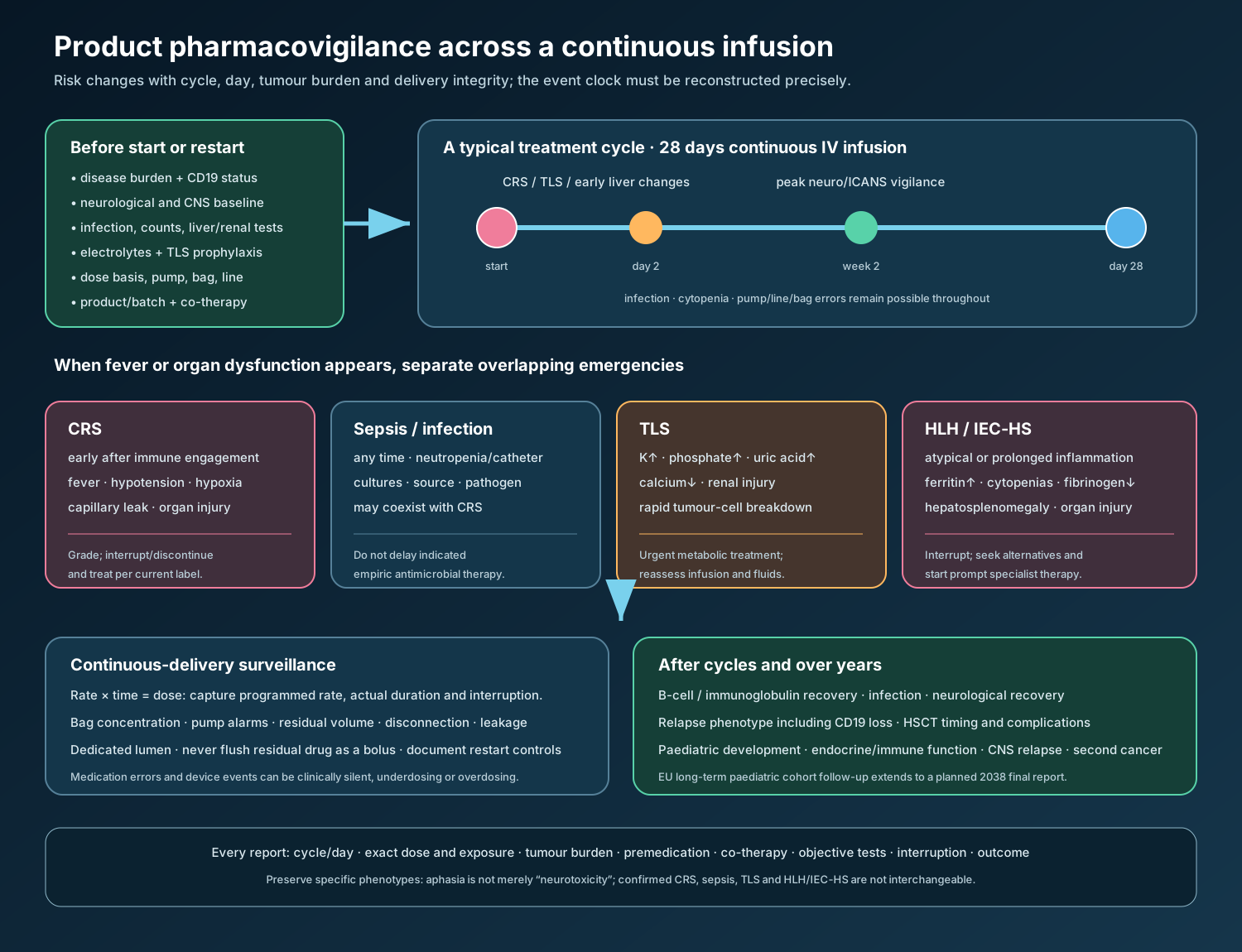
Before every course: verify indication, CD19 status where clinically relevant, disease burden, prior therapy/HSCT, CNS and neurological history, infection, blood counts, liver and renal function, electrolytes, immunoglobulins where appropriate, pregnancy considerations, intrathecal plan, exact dose basis, pump programme, product and batch.
First 48 hours: give particular attention to CRS, infusion reaction, TLS, hypotension, capillary leak, fever, early hepatic changes, and preparation or rate errors. Document dexamethasone, cytoreduction, hydration and anti-hyperuricaemic prophylaxis.
First two to three weeks: maintain structured neurological assessment for tremor, writing or language change, inattention, confusion, ataxia, seizure and ICANS. Neurological events can occur later, so this is a peak window rather than a stop date.
Entire continuous infusion: monitor fever and infection, cytopenias, catheter and pump integrity, bag changes, occlusion/disconnection, interruptions, accidental bolus, underdose, overdose, and home-care escalation. A dose received is a rate multiplied by time; both variables must be reconstructed in a medication-error report.
After cycles and long term: assess B-cell and immunoglobulin recovery, infection, relapse phenotype including CD19 loss, transplant outcome, neurological recovery, and paediatric development/secondary malignancy as applicable.
Differentiating four inflammatory emergencies
| Feature | CRS | Sepsis/infection | TLS | HLH/IEC-HS |
|---|---|---|---|---|
| Typical clue | Starts soon after T-cell engagement; day 2 peak in pooled data | Pathogen/source or neutropenic context; can occur anytime | Biochemical cell-breakdown pattern after rapid response | Persistent/atypical hyperinflammation, often after or overlapping CRS |
| Core findings | Fever, hypotension, hypoxia, capillary leak, organ dysfunction | Fever/hypothermia, hypotension, focal or culture evidence, organ dysfunction | High K/phosphate/uric acid, low Ca, acute kidney injury | Very high ferritin, cytopenias, low fibrinogen, liver/spleen enlargement, hepatitis |
| Dangerous assumption | “All fever is infection” | “All fever after infusion is CRS” | “Electrolytes are just chemotherapy effects” | “Prolonged CRS needs only more observation” |
| Immediate logic | Grade, interrupt when required, supportive/corticosteroid or cytokine-directed care per protocol | Cultures and prompt empiric treatment when indicated | Stop/reassess therapy, fluids and urgent metabolic management | Interrupt; diagnostic work-up for infection/malignancy/autoimmunity; prompt specialist therapy |
These diagnoses can coexist. The table organizes reasoning; it is not a bedside diagnostic rule.
Medication-error and device-event capture
Continuous microgram dosing turns small process failures into clinically important exposure changes. A useful report includes:
- prescribed and intended dose, weight/body-surface-area basis, prepared concentration, diluent and bag duration;
- pump make/model, programmed rate, start/stop times, alarms, residual volume, occlusion, disconnection, leakage or power failure;
- whether the dedicated line was flushed, clamped, primed, paused, changed, or used for another medicine;
- estimated actual dose and uncertainty, symptoms, tests, treatment, outcome and re-exposure;
- preparation site, personnel roles, instructions used, handovers, home-care context, product name and batch.
Device malfunction may require reporting under medical-device as well as medicinal-product systems. A near miss without patient exposure remains valuable for root-cause prevention.
Case assessment, coding and signal detection
Retain reporter language and code the most specific supported concepts. “Neurological toxicity” should not erase aphasia, dysgraphia, seizure, encephalopathy, or ICANS. “Infusion reaction” should not absorb confirmed CRS, TLS, sepsis, or HLH/IEC-HS. Seriousness follows regulatory outcome criteria, while severity describes intensity.
Causality assessment should consider cycle/day, interruption and restart, tumour burden, concurrent chemotherapy, HSCT, infection, CNS leukaemia, electrolyte disturbance, corticosteroids and other neurotoxic medicines. Rapid improvement after stopping a two-hour-half-life medicine supports—but does not prove—causality.
Aggregate surveillance should stratify CRS and ICANS by indication, age, cycle, dose step, tumour burden, inpatient versus home setting, and concomitant therapy. Search for disproportionate severity, late onset, recurrence on restart, fatal outcome, pump pattern, and product/batch clustering rather than counting undifferentiated events.
Evolution of the safety system
The 2014 US accelerated approval was based on a single-arm relapsed/refractory study and introduced a REMS communication plan for CRS, neurological toxicity, and preparation/administration errors.[4,11] The programme uses education rather than restricted distribution or prescriber/patient certification. Later randomized evidence confirmed benefit, indications expanded, and the REMS materials were modified to match populations and risk-management knowledge.[5,11]
European conditional authorisation in 2015 became standard authorisation in 2018 after confirmatory TOWER data.[1,12] Initial EU educational materials emphasized neurological events and medication errors. By RMP version 19.0, medication errors had been removed from the formal important-risk list and educational materials for pharmacists and physicians were no longer considered necessary; nurse and patient/caregiver materials and a patient card remained, with the card addressing ICANS and CRS.[3]
Safety terminology also evolved. ICANS was added explicitly as knowledge of immune-effector neurotoxicity matured. The 2025 RMP changed “neurologic events” to “neurologic events including ICANS,” based on clinical and postmarketing observations.[3] The January 2026 EU information added an HLH/IEC-HS warning and management instructions.[1] These changes show why historical labels should not be used as current reference safety information.
Long-term paediatric surveillance is scheduled over decades in the EU RMP, including a cohort study with a final report planned for 2038.[3] Such follow-up is necessary because survival benefit can make late development, immune recovery, relapse patterns and second malignancies observable only years later.
What the medicine solves—and what it does not
Blinatumomab can convert MRD-positive remission to MRD negativity, improve response and survival in relapsed/refractory disease, and improve survival when added to consolidation in selected newly diagnosed Philadelphia-negative disease. It can reduce reliance on another block of intensive chemotherapy in some protocols.
It does not guarantee cure, treat CD19-negative disease, replace CNS prophylaxis, directly inhibit BCR::ABL1, eliminate transplant decisions, or remove the need for multi-agent therapy in newly diagnosed ALL. The same T-cell bridge that supplies benefit creates a time-sensitive safety system extending from the pharmacy clean room to the patient’s home.
Official regulatory reference material
Use the latest jurisdiction-specific versions; the following primary materials were checked on 3 September 2026.
- EMA EPAR and current Product Information (SmPC and package leaflet)—indications, dosing, contraindications, warnings, adverse reactions and January 2026 HLH/IEC-HS update.
- EMA EU Risk Management Plan, version 19.0 (9 April 2025)—current published safety concerns, pharmacovigilance plan and additional risk-minimisation measures.
- FDA US Prescribing Information, BLA 125557—boxed warning, labelled indications, dosing, Instructions for Use and Medication Guide.
- FDA approval packages and multidisciplinary reviews—original 2014 approval, 2017 TOWER confirmation, 2018 MRD expansion and 2024 consolidation expansion.
- FDA BLINCYTO REMS document and modification letters—communication-plan goals and materials.
- EMA EPAR assessment reports and post-authorisation variation assessments—development history and evolution of authorisation.
References
- European Medicines Agency. Blinatumomab EPAR and current product information. Product information updated 15 January 2026.
- US Food and Drug Administration. Blinatumomab US Prescribing Information, BLA 125557/S-032. 21 October 2025.
- European Medicines Agency. Blinatumomab EU Risk Management Plan, version 19.0. 9 April 2025.
- US Food and Drug Administration. Original clinical review: blinatumomab, BLA 125557. 2014.
- Kantarjian H, et al. Blinatumomab versus chemotherapy for advanced acute lymphoblastic leukemia. N Engl J Med. 2017;376:836–847.
- US Food and Drug Administration. FDA supplemental-approval analysis of TOWER and ALCANTARA. Oncologist. 2018;23:1366–1371.
- Gökbuget N, et al. Blinatumomab for minimal residual disease in adults with B-cell precursor acute lymphoblastic leukemia. Blood. 2018;131:1522–1531.
- US Food and Drug Administration. 2018 approval for MRD-positive B-cell precursor ALL. 29 March 2018.
- Litzow MR, et al. Blinatumomab for MRD-negative acute lymphoblastic leukemia in adults. N Engl J Med. 2024;391:320–333.
- US Food and Drug Administration. 2024 approval as consolidation for CD19-positive Philadelphia-negative BCP-ALL. 14 June 2024.
- US Food and Drug Administration. Blinatumomab REMS document. Current programme and modification history should be checked in REMS@FDA.
- European Medicines Agency. Blinatumomab initial EPAR public assessment report. 2015.
- Bargou R, et al. Tumor regression in cancer patients by very low doses of a T cell-engaging antibody. Science. 2008;321:974–977.
- Goebeler ME, Bargou RC. T cell-engaging therapies—BiTEs and beyond. Nat Rev Clin Oncol. 2020;17:418–434.
Regulatory Note
This article is educational and does not replace the current SmPC, package leaflet, US Prescribing Information, Instructions for Use, Medication Guide, RMP, REMS, institutional protocol, or clinical judgement. Indications and risk-minimisation requirements differ between jurisdictions and can change. Treatment must be initiated and supervised by clinicians experienced in acute lymphoblastic leukaemia and continuous infusion. Suspected adverse reactions, medication errors, device problems, and quality defects should be reported through the applicable systems, with exact product and batch traceability whenever available.