Brolucizumab: Classification, History, Mechanism of Action, Safety and Pharmacovigilance
Brolucizumab is an intravitreal single-chain antibody fragment (scFv) that binds vascular endothelial growth factor A (VEGF-A). It was designed to deliver a high molar amount of VEGF-binding capacity in a small protein format. By neutralising VEGF-A within the eye, it reduces pathological vascular permeability and neovascular activity in retinal disease.
Its pharmacovigilance is dominated by a different problem from most systemic monoclonal antibodies: the clinically important event often occurs in one treated eye, and the differential diagnosis includes the molecule, the injection procedure, retinal vascular pathology and progression of the underlying disease. A report of “visual loss” is therefore not a sufficiently specific safety case. Laterality, ocular examination, imaging and injection chronology are central evidence.
- Brolucizumab: Classification, History, Mechanism of Action, Safety and Pharmacovigilance
- Multidimensional classification
- VEGF-A and retinal vascular disease
- Mechanism of action
- Development and regulatory history
- Clinical use and treatment-response context
- Major safety domains
- Anti-drug antibodies and immunogenicity
- Product identification, laterality and bilateral treatment
- Pharmacovigilance case assessment
- Signal detection and aggregate review
- Periodic benefit-risk evaluation
- Risk management and operational controls
- Potential failure modes
- Inspection and governance perspective
- Practical checklist
- Key Takeaways
- References
- Regulatory Note
Multidimensional classification
| Classification axis | Brolucizumab classification | Scientific or PV significance |
|---|---|---|
| Molecular format | Humanised single-chain variable antibody fragment | Much smaller than a full IgG and lacks a conventional Fc region |
| Target | VEGF-A | Inhibits a central mediator of vascular permeability and pathological angiogenesis |
| Therapeutic class | Intravitreal anti-VEGF biological | Safety includes both molecule-related and procedure-related ocular events |
| Current EU indications | Neovascular age-related macular degeneration and visual impairment due to diabetic macular oedema | Background ocular and systemic vascular risks differ by indication |
| Route | Intravitreal injection | Laterality, aseptic procedure and injection chronology are essential PV variables |
| Major identified ocular concerns | Intraocular inflammation; retinal vasculitis and/or retinal vascular occlusion; endophthalmitis; IOP increase; retinal tear/detachment | Requires examination-level case follow-up |
| Systemic consideration | VEGF-pathway exposure after ocular dosing | Systemic vascular events need contextual assessment rather than temporal attribution alone |
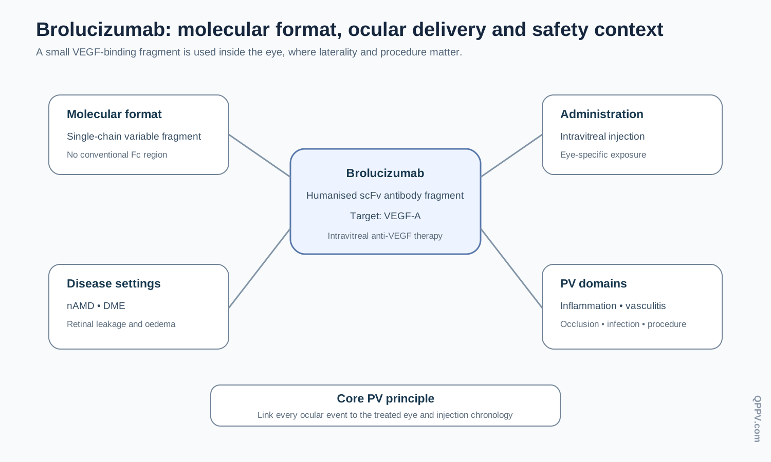
Figure 1. Brolucizumab combines a small anti-VEGF antibody-fragment format with intravitreal delivery. Pharmacovigilance must separate target pharmacology, ocular inflammation, injection complications and underlying retinal disease.
VEGF-A and retinal vascular disease
VEGF-A promotes endothelial permeability, pathological new-vessel formation and leakage. In neovascular age-related macular degeneration (nAMD), abnormal choroidal vessels can leak fluid or blood beneath and within the retina. In diabetic macular oedema (DME), retinal vascular dysfunction and permeability contribute to accumulation of fluid in the macula.
Neutralising VEGF-A reduces leakage and pathological angiogenic signalling. This mechanism is shared with other intravitreal anti-VEGF therapies, but molecular architecture, dose, pharmacokinetics and product-specific safety evidence differ between products.
Why the antibody-fragment format matters
Brolucizumab consists of linked variable antibody domains rather than a full-length IgG. The small molecular size permits a high molar concentration of VEGF-binding sites within the limited injection volume of the eye. That design feature is relevant to pharmacology but should not be used to infer safety without evidence; product-specific post-authorisation data remain decisive.
Mechanism of action
Brolucizumab binds VEGF-A and prevents VEGF-A from activating VEGF receptors on endothelial cells. The downstream result is reduced vascular permeability, reduced pathological angiogenic signalling and, in responsive eyes, reduction of retinal or subretinal fluid.
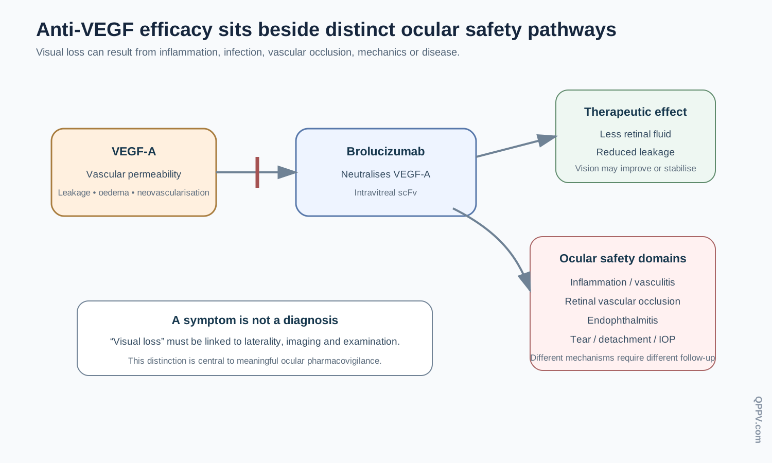
Figure 2. VEGF-A neutralisation reduces retinal vascular leakage, while intravitreal treatment introduces distinct inflammatory, infectious and mechanical safety pathways. Visual deterioration must therefore be phenotyped rather than treated as one event category.
Development and regulatory history
The European Union authorised brolucizumab in February 2020 for neovascular AMD. The authorised use later expanded to include visual impairment due to diabetic macular oedema.
Post-authorisation experience materially changed the safety understanding. Reports of intraocular inflammation, including retinal vasculitis and retinal vascular occlusion, led to strengthened warnings, risk-minimisation measures and direct communications to healthcare professionals. EMA risk-management documentation now treats retinal vasculitis and/or retinal vascular occlusion as an important identified risk and notes that severe vision loss has occurred in some post-marketing cases.
This history is a useful example of pharmacovigilance changing product use after approval. The relevant lesson is not that pre-authorisation trials were irrelevant; it is that rare, clinically distinctive ocular events may become clearer only after larger and more heterogeneous real-world exposure.
Clinical use and treatment-response context
Brolucizumab is administered by intravitreal injection into the affected eye. Treatment begins with a loading phase and then moves to longer intervals according to the authorised regimen and disease activity. Exposure therefore cannot be reconstructed from the statement “receives anti-VEGF injections” alone. The actual eye, injection date, interval, previous anti-VEGF therapy and whether treatment was bilateral are relevant.
The indication also changes background risk. Patients with nAMD are often older and may have substantial cardiovascular comorbidity. Patients with DME commonly have diabetes, hypertension and systemic vascular disease. Those baseline risks matter when interpreting stroke, myocardial infarction or retinal vascular events.
Major safety domains
Intraocular inflammation
Intraocular inflammation can range from anterior chamber inflammation to vitritis and more severe posterior-segment inflammatory disease. In brolucizumab pharmacovigilance, inflammation is particularly important because retinal vasculitis and retinal vascular occlusion have often occurred in the presence of intraocular inflammation.
A case should capture laterality, onset after injection, anterior- and posterior-segment findings, visual acuity, optical coherence tomography, fundus examination, angiography where performed, treatment and outcome. A generic term such as “uveitis” is less useful when the anatomical phenotype is known.
Retinal vasculitis and retinal vascular occlusion
Post-authorisation reports established retinal vasculitis and/or retinal vascular occlusion as important identified risks. Some cases have been associated with severe visual loss. EMA risk-management material notes that a history of intraocular inflammation or retinal vascular occlusion may identify a higher-risk population and recommends careful examination before treatment and discontinuation if retinal vasculitis or retinal vascular occlusion develops.
The distinction between vasculitis and vascular occlusion matters. Vasculitis describes inflammatory involvement of retinal vessels; occlusion describes impaired blood flow and can be arterial or venous. They can coexist, but a high-quality case should record which diagnosis was made and on what evidence.
Endophthalmitis
Endophthalmitis is a recognised risk of intravitreal injection. It may present with pain, redness, hypopyon, vitritis and rapid visual decline. Its occurrence after brolucizumab does not automatically indicate a molecule-specific immune reaction.
Follow-up should include aseptic-procedure details, onset, microbiology, intravitreal antimicrobial treatment, vitrectomy if performed and visual outcome. Sterile inflammation and infectious endophthalmitis require different clinical and pharmacovigilance interpretation.
Retinal tear and retinal detachment
Intravitreal procedures can be followed by retinal tears or detachment. These events are generally procedure-related or disease-related rather than direct consequences of VEGF neutralisation. The safety case should include retinal location, timing, posterior-vitreous status, procedure details, surgery and outcome.
Intraocular pressure
Transient intraocular-pressure elevation can occur after intravitreal injection. Sustained pressure changes require assessment of baseline glaucoma or ocular hypertension, serial measurements, optic-nerve status and treatment. A pressure event should not be interpreted without knowing whether it occurred immediately after injection or developed over longer follow-up.
Systemic arterial thromboembolic events
VEGF has physiological vascular functions, so arterial thromboembolic events remain a class-relevant question for intravitreal anti-VEGF therapy. However, the treated population already has substantial baseline vascular risk.
A stroke or myocardial infarction after injection therefore requires reconstruction of diabetes, hypertension, atrial fibrillation, prior vascular disease, bilateral treatment, recent injections and competing causes. Temporal proximity alone does not establish causation.
Anti-drug antibodies and immunogenicity
As a therapeutic protein, brolucizumab can induce anti-drug antibodies. Immunogenicity is particularly relevant when investigating intraocular inflammation, but antibody positivity alone does not prove that an inflammatory event was caused by anti-drug antibodies.
Useful cases link immunogenicity results with timing, ocular phenotype, prior injections and recurrence. Aggregate analyses should avoid using anti-drug-antibody positivity as a substitute for clinical diagnosis.
Product identification, laterality and bilateral treatment
The exposure record should preserve the exact product, batch, injected eye and injection date. Bilateral treatment creates a natural within-patient comparison but also complicates attribution if both eyes are exposed on different dates or to different anti-VEGF products.
Laterality is therefore a core exposure field, not an optional ophthalmology detail. A system unable to link the event eye to the injected eye loses essential causal information.
Pharmacovigilance case assessment
Brolucizumab cases should be reconstructed around four core variables: treated eye, injection chronology, ocular phenotype and indication. Visual symptoms become interpretable only when those variables are linked to objective examination or imaging.
Event-specific follow-up priorities
| Event or issue | High-value follow-up information |
|---|---|
| Intraocular inflammation | Laterality, onset, anterior/vitreous findings, visual acuity, OCT, treatment and recurrence |
| Retinal vasculitis | Angiography, arterial/venous involvement, occlusive status, inflammation, visual change and outcome |
| Retinal vascular occlusion | Vessel involved, imaging, inflammatory findings, baseline vascular disease and treatment |
| Endophthalmitis | Injection date, asepsis, pain/redness, hypopyon/vitritis, microbiology, intravitreal therapy, vitrectomy and outcome |
| Retinal tear/detachment | Timing, retinal location, procedure details, surgery and final vision |
| IOP increase | Baseline IOP/glaucoma, post-dose measurements, duration, treatment and optic-nerve findings |
| Lack of efficacy | Indication, visual acuity, serial OCT, injection interval, missed visits and prior anti-VEGF therapy |
| Systemic vascular event | Event phenotype, vascular risk factors, bilateral treatment, injection timing and competing causes |
Signal detection and aggregate review
Retinal vasculitis and retinal vascular occlusion should be analysed as dedicated medically coherent case series, with inflammatory phenotype preserved. Combining them indiscriminately with all ocular inflammation or all retinal vascular events can obscure the specific pattern that prompted regulatory action.
Endophthalmitis should remain separate from sterile intraocular inflammation. Both can produce pain, redness and visual loss, but their mechanisms, treatment and regulatory meaning differ. Aggregate retrieval should therefore support broad screening while preserving clinically specific diagnoses for medical review.
Analyses should also stratify by indication and prior anti-VEGF treatment. Patients with DME differ from those with nAMD in age, diabetic vascular disease and retinal pathology. Prior intraocular inflammation or vascular occlusion is particularly relevant to risk assessment.
Periodic benefit-risk evaluation
Periodic evaluation should integrate anatomical and visual benefit with intraocular inflammation, retinal vasculitis/occlusion, endophthalmitis, retinal complications, intraocular pressure, immunogenicity and systemic vascular observations.
The post-authorisation history demonstrates why benefit-risk assessment is longitudinal. A medicine may retain clinically meaningful efficacy while new information changes which patients should receive it, how they are monitored and what symptoms require urgent evaluation.
Risk management and operational controls
Current product information and risk-minimisation materials govern examination before treatment, contraindications, recognition of intraocular inflammation and management of retinal vasculitis or vascular occlusion. Educational materials for patients are part of the EU risk-minimisation framework.
Recommended operational controls include mandatory laterality fields, structured ocular follow-up, separation of infectious versus sterile inflammation, targeted retinal-vasculitis review and product/batch capture. These operational controls support implementation; they should not be presented as legal requirements unless specifically mandated.
Potential failure modes
The following are illustrative scenarios, not published inspection findings:
- Visual loss is coded without identifying the treated eye or ocular diagnosis.
- Endophthalmitis and sterile intraocular inflammation are pooled into one signal.
- Retinal vasculitis is recorded without angiography or occlusive status where these were available.
- A retinal tear is attributed solely to VEGF pharmacology without considering the injection procedure.
- Prior intraocular inflammation is omitted from a retinal vascular-occlusion case.
- Apparent treatment failure is assessed without reconstructing injection intervals, missed visits or serial OCT findings.
Inspection and governance perspective
An inspector assessing brolucizumab pharmacovigilance could examine whether serious ocular cases retain laterality, objective examination and imaging data; whether retinal vasculitis/occlusion receives targeted medical review; whether the organisation can distinguish molecule-related inflammation from infectious and mechanical complications; and whether risk-minimisation communications are reflected in operational follow-up.
The effectiveness question is whether the safety system can recognise the specific post-authorisation ocular phenotype that changed the product’s risk management, rather than merely count generic terms such as “visual impairment” or “uveitis”.
Practical checklist
For a brolucizumab case or aggregate analysis, confirm:
- indication and treated eye;
- exact injection date and treatment interval;
- prior anti-VEGF treatment and ocular inflammatory history;
- visual acuity and objective ophthalmic findings;
- OCT and angiography where relevant;
- distinction between sterile inflammation and infection;
- vasculitis and vascular-occlusion phenotype;
- IOP measurements for pressure events;
- procedure details for mechanical complications;
- systemic vascular risk factors for stroke or myocardial infarction;
- exact product, batch and administration details.
Key Takeaways
Brolucizumab is a humanised single-chain antibody fragment that neutralises VEGF-A inside the eye. Its small format permits high molar VEGF-binding capacity, but product-specific safety evidence—not molecular size alone—determines its benefit-risk profile.
Its pharmacovigilance is dominated by intraocular inflammation, retinal vasculitis and retinal vascular occlusion, alongside the established procedural risks of intravitreal therapy. Laterality, imaging and injection chronology are therefore essential to meaningful case assessment.
References
- European Medicines Agency. Brolucizumab: EPAR and current product information. EU marketing authorisation issued February 2020. https://www.ema.europa.eu/en/medicines/human/EPAR/beovu
- European Medicines Agency. Brolucizumab product information. https://www.ema.europa.eu/en/documents/product-information/beovu-epar-product-information_en.pdf
- European Medicines Agency. Brolucizumab EU risk-management plan. Retinal vasculitis and/or retinal vascular occlusion are important identified risks. https://www.ema.europa.eu/en/documents/rmp/beovu-epar-risk-management-plan_en.pdf
- European Medicines Agency. Direct healthcare professional communication: updated recommendations to minimise intraocular inflammation including retinal vasculitis and/or retinal vascular occlusion. 2021. https://www.ema.europa.eu/en/medicines/dhpc/beovu
- Dugel PU, Koh A, Ogura Y, et al. HAWK and HARRIER: phase 3, multicenter, randomized, double-masked trials of brolucizumab for neovascular age-related macular degeneration. Ophthalmology. 2020;127:72-84. doi:10.1016/j.ophtha.2019.04.017.
Regulatory Note
Authorised indications, injection intervals, contraindications and ocular safety recommendations can change and differ between regions. This article explains the scientific and pharmacovigilance framework and does not replace current regional product information or specialist retinal guidance. Regulatory information was checked against EMA material current in September 2026.