Faricimab: Classification, History, Mechanism of Action, Safety and Pharmacovigilance
- Faricimab: Classification, History, Mechanism of Action, Safety and Pharmacovigilance
- Multidimensional classification
- Why VEGF-A and Ang-2 are paired targets
- Molecular engineering and mechanism
- Development and regulatory history
- Clinical use and treatment-response context
- Major safety domains
- Anti-drug antibodies and immunogenicity
- Product identification and bilateral treatment
- Pharmacovigilance case assessment
- Signal detection and aggregate review
- Periodic benefit-risk evaluation
- Risk management and operational controls
- Potential failure modes
- Inspection and governance perspective
- Practical checklist
- Key Takeaways
- References
- Regulatory Note
Faricimab is a bispecific antibody designed for intravitreal treatment of retinal vascular disease. One binding arm neutralises vascular endothelial growth factor A (VEGF-A), while the other binds angiopoietin-2 (Ang-2). The therapeutic idea is not simply to add a second target to anti-VEGF therapy. VEGF-A and Ang-2 influence overlapping aspects of vascular permeability, endothelial survival, inflammation and vessel stability, so dual inhibition is intended to address both active leakage and the vascular context in which leakage persists.
The pharmacovigilance problem is correspondingly multidimensional. An event after faricimab may reflect drug pharmacology, an immune-inflammatory response, the intravitreal procedure, pre-existing retinal disease, or progression despite treatment. Sudden visual loss, for example, can arise from endophthalmitis, retinal vascular occlusion, retinal tear or detachment, severe intraocular inflammation, haemorrhage or the underlying macular disease. High-quality PV therefore depends on ophthalmic phenotype and examination findings rather than on the symptom term alone.
Multidimensional classification
| Classification axis | Faricimab classification | Scientific or PV significance |
|---|---|---|
| Molecular class | Bispecific monoclonal antibody | Two antigen-binding specificities are incorporated into one molecule |
| Targets | VEGF-A and Ang-2 | Links anti-permeability/anti-angiogenic activity with modulation of vascular stability |
| Therapeutic class | Intravitreal retinal vascular therapy | Local ocular administration changes exposure and adverse-event attribution |
| Disease context | nAMD, DME and macular oedema due to retinal vein occlusion in the EU | Baseline retinal pathology differs between indications |
| Procedure | Intravitreal injection | Introduces procedure-related risks independent of antibody pharmacology |
| PV-critical ocular risks | Intraocular inflammation, retinal vasculitis/vascular occlusion, endophthalmitis, retinal tear/detachment, IOP change | Requires examination-level follow-up and laterality |
| Systemic consideration | VEGF-pathway exposure after ocular dosing | Systemic exposure is lower than ocular exposure but vascular events require contextual assessment |
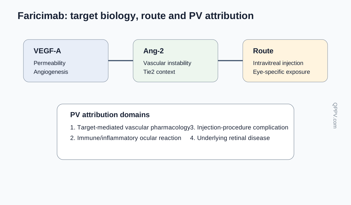
Figure 1. Faricimab combines a dual-target vascular mechanism with intravitreal delivery. Pharmacovigilance must therefore separate target-mediated effects, ocular inflammation, injection-procedure complications and disease progression.
Why VEGF-A and Ang-2 are paired targets
VEGF-A is a central driver of pathological vascular permeability and neovascularisation in the retina. It increases endothelial permeability, promotes vascular growth and contributes to fluid accumulation in macular disease. Inhibiting VEGF-A therefore reduces leakage and pathological new-vessel activity.
Angiopoietin signalling acts through the endothelial receptor Tie2. Angiopoietin-1 generally supports vascular quiescence and endothelial stability. Ang-2 can antagonise this stabilising environment and, particularly in the presence of inflammatory or VEGF signals, promote vascular destabilisation and permeability. The simplified teaching model is therefore that VEGF-A drives leakage and angiogenesis while Ang-2 reduces vascular stability; in biological systems the pathways are more interdependent than that shorthand suggests.
Dual inhibition is not two independent drugs
Because both binding specificities are carried by the same molecule, ocular exposure, pharmacokinetics, immunogenicity and product quality belong to one biological product. Signal assessment should not try to assign an ocular inflammatory event to the “VEGF arm” or “Ang-2 arm” unless evidence genuinely supports such a distinction.
Molecular engineering and mechanism
Faricimab uses an engineered bispecific antibody architecture that permits two different antigen-binding arms while retaining an antibody-like molecular format. Fc-region engineering is used to minimise unwanted Fc-mediated effector interactions, because retinal efficacy depends on ligand neutralisation rather than immune-cell recruitment.
By binding VEGF-A, faricimab reduces VEGF-receptor activation. By binding Ang-2, it reduces Ang-2-mediated interference with Tie2-associated vascular stability. The intended downstream result is reduced leakage, reduced pathological neovascular activity and a vascular environment capable of remaining controlled for longer treatment intervals in some patients.
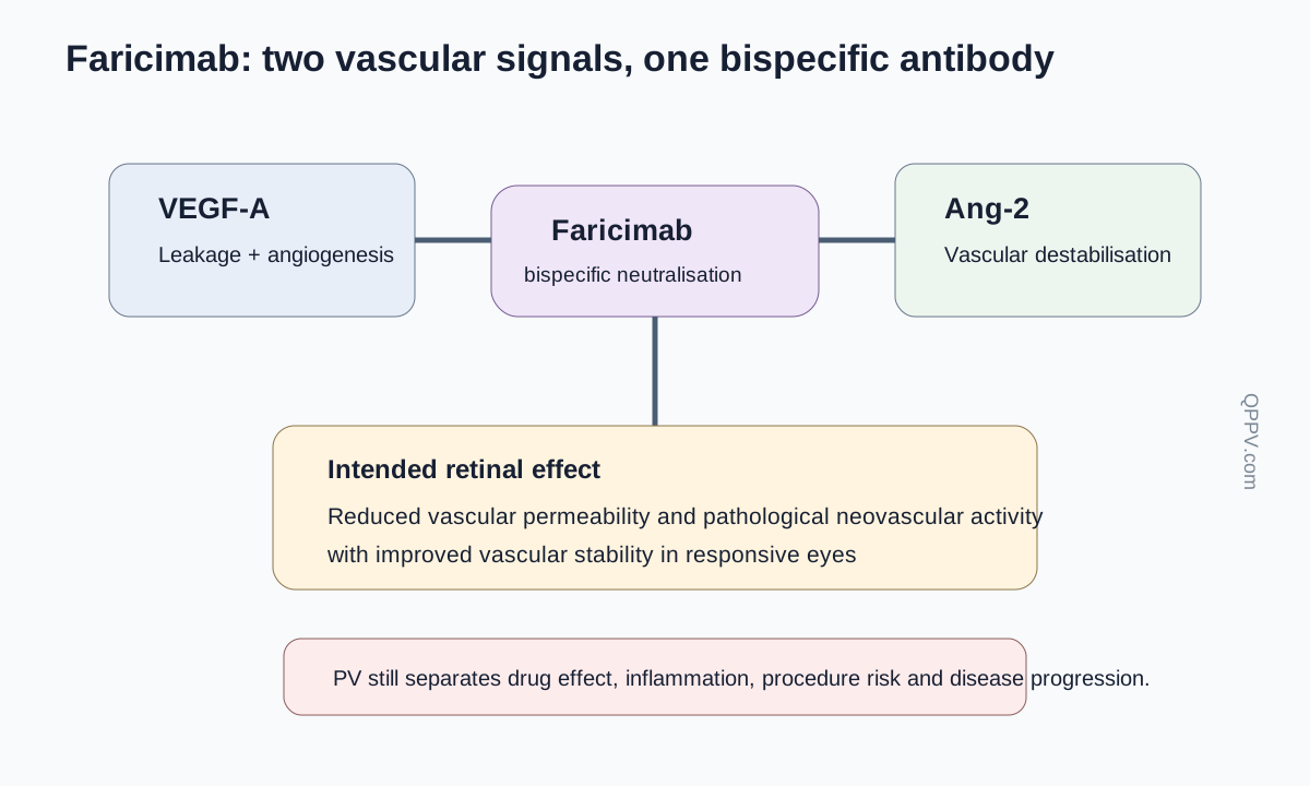
Figure 2. VEGF-A promotes permeability and pathological angiogenesis, while Ang-2 can destabilise Tie2-mediated vascular quiescence. Faricimab neutralises both ligands. The pathways overlap, so the figure represents complementary vascular mechanisms rather than two isolated effects.
Development and regulatory history
Faricimab was developed after anti-VEGF therapy had already transformed management of neovascular age-related macular degeneration and diabetic macular oedema. The remaining clinical challenge included treatment burden: effective VEGF suppression often required repeated intravitreal injections and monitoring. Development therefore examined whether combined VEGF-A and Ang-2 inhibition could preserve visual and anatomical outcomes while allowing longer intervals in appropriately responsive patients.
The European Union authorised faricimab in 2022 for neovascular age-related macular degeneration and visual impairment due to diabetic macular oedema. The EU indication subsequently expanded to visual impairment due to macular oedema secondary to retinal vein occlusion. Current EMA product information was updated in August 2026, so exact dosing and safety wording should be checked against the current document.
Clinical use and treatment-response context
Faricimab is administered by intravitreal injection. The relevant treatment interval is determined by the authorised regimen and the individual response of the treated eye. This means that exposure cannot be interpreted simply as “monthly” or “every few months” without knowing the treatment phase, disease activity, interval extension history and whether both eyes are treated.
The treated indication changes the competing explanations for visual symptoms. Neovascular age-related macular degeneration can produce haemorrhage, fibrosis and recurrent exudation. Diabetic macular oedema occurs in a population with systemic vascular disease and other diabetic retinopathy manifestations. Retinal vein occlusion carries its own vascular and haemorrhagic background. Aggregate analyses should therefore preserve indication and laterality.
Major safety domains
Intraocular inflammation and retinal vasculitis
Intraocular inflammation can range from anterior chamber cells or vitritis to more severe inflammatory syndromes. Post-authorisation reports of retinal vasculitis and retinal vascular occlusion have made precise ophthalmic characterisation especially important. A case should record whether vasculitis was occlusive or non-occlusive, arterial or venous involvement where known, laterality, imaging findings, visual-acuity change, treatment and outcome.
Inflammation should not be collapsed into “uveitis” when the actual phenotype is known. The distinction between sterile inflammation, infectious endophthalmitis and retinal vascular inflammation changes both causality and clinical urgency.
Endophthalmitis
Endophthalmitis is a recognised risk of intravitreal injection. Its occurrence after faricimab does not automatically imply a molecule-specific immune reaction. High-value follow-up includes injection date, aseptic procedure details, onset, pain, hypopyon, vitreous findings, culture or PCR, intravitreal antibiotics, surgery and outcome.
Retinal tear, retinal detachment and traumatic lens injury
Intravitreal procedures can be followed by retinal tear or detachment, and incorrect needle placement can damage intraocular structures. These events are best treated as procedure-related safety problems unless evidence suggests another mechanism. Device or administration information may therefore be as important as product exposure.
Intraocular pressure
Transient increases in intraocular pressure can occur after intravitreal injection, and sustained pressure changes may have multiple causes. Cases should include baseline glaucoma or ocular hypertension, pre- and post-injection pressure, timing, optic-nerve status and treatment.
Systemic arterial thromboembolic events
VEGF has physiological vascular functions, so systemic arterial thromboembolic events remain a class-relevant question for intravitreal anti-VEGF therapies. However, retinal populations commonly have advanced age, diabetes, hypertension and established vascular disease. A myocardial infarction or stroke after injection therefore requires careful background-risk assessment rather than temporal attribution alone.
Anti-drug antibodies and immunogenicity
As with other therapeutic proteins, anti-drug antibodies can develop. Their clinical importance depends on whether they alter ocular exposure, efficacy or inflammatory risk. An inflammatory event accompanied by anti-drug antibodies may be mechanistically informative, but antibody positivity alone does not prove causation.
Product identification and bilateral treatment
PV systems should preserve the exact product, batch, treated eye, injection date and whether the fellow eye received the same or another anti-VEGF therapy. Bilateral treatment can complicate attribution if an ocular event occurs in only one eye. Laterality is therefore not optional clinical detail; it is part of the exposure record.
Pharmacovigilance case assessment
Faricimab cases should be reconstructed around four variables: treated eye, indication, injection chronology and objective ophthalmic findings. Visual loss without these variables is a symptom report, not a clinically interpretable ocular safety case.
Event-specific follow-up priorities
| Event | High-value follow-up information |
|---|---|
| Intraocular inflammation | Laterality, onset, anterior/vitreous findings, OCT, fundus findings, treatment, recurrence, anti-drug antibodies if available |
| Retinal vasculitis/occlusion | Angiography/OCT findings, arterial/venous territory, occlusive status, visual change, inflammation, treatment and outcome |
| Endophthalmitis | Injection date, asepsis, pain/redness, hypopyon/vitritis, microbiology, intravitreal antibiotics, vitrectomy, outcome |
| Retinal tear/detachment | Timing, retinal location, posterior vitreous status, procedure details, surgery and visual outcome |
| IOP increase | Baseline IOP/glaucoma, post-dose measurements, duration, treatment, optic-nerve findings |
| Lack of efficacy | Indication, baseline and serial OCT, visual acuity, interval history, missed visits, prior anti-VEGF therapy |
| Systemic vascular event | Stroke/MI phenotype, vascular risk factors, bilateral treatment, recent injections, competing causes |
Signal detection and aggregate review
Ocular signal detection should separate inflammatory events from infectious and mechanical procedural complications. Retinal vasculitis warrants its own medically coherent case series rather than being pooled indiscriminately with all uveitis terms. Analyses should also preserve indication, laterality, treatment interval and prior intravitreal therapy.
When comparing products, spontaneous-report counts should not be treated as incidence. Differences in market exposure, patient mix, reporting intensity, launch timing and diagnostic awareness can all alter observed reporting patterns.
Periodic benefit-risk evaluation
Periodic review should integrate visual and anatomical benefit with serious ocular risks, treatment burden, systemic vascular observations, immunogenicity and administration problems. Extension of treatment intervals is clinically valuable only if disease control is maintained; therefore reports of recurrence or visual deterioration should be interpreted in relation to interval history rather than as generic lack-of-efficacy cases.
Risk management and operational controls
Current product information governs intravitreal administration, contraindications, ocular monitoring and management of inflammatory or infectious complications. Useful PV controls include structured ophthalmology follow-up, mandatory laterality fields, separate coding conventions for endophthalmitis versus sterile inflammation, retinal-vasculitis case review and product/batch capture.
Potential failure modes
The following are illustrative scenarios, not published inspection findings:
- Sudden visual loss is coded without identifying the treated eye or ophthalmic diagnosis.
- Endophthalmitis and sterile intraocular inflammation are pooled as one signal.
- Retinal vasculitis is recorded without angiographic or occlusive-status information.
- A procedure-related retinal tear is attributed solely to VEGF/Ang-2 pharmacology.
- Apparent treatment failure is assessed without reconstructing injection intervals or missed visits.
- Bilateral treatment is not captured, making eye-specific exposure impossible to reconstruct.
Inspection and governance perspective
An inspector assessing faricimab pharmacovigilance could examine whether ocular cases retain laterality, procedural chronology and objective ophthalmic evidence; whether serious inflammation and vasculitis receive targeted medical review; and whether product-quality, device and batch information can be linked to clusters. The effectiveness question is whether the system can distinguish the pharmacology of the antibody from the hazards of delivering any medicine into the vitreous cavity.
Practical checklist
For a faricimab case or aggregate analysis, confirm:
- indication and treated eye;
- exact injection date and treatment interval;
- prior and concomitant intravitreal therapies;
- visual acuity and objective ocular examination findings;
- OCT/angiography or microbiology where relevant;
- distinction between inflammatory, infectious, vascular and mechanical diagnoses;
- IOP trajectory for pressure events;
- systemic vascular risk factors for stroke or myocardial infarction;
- product, batch and administration details.
Key Takeaways
Faricimab is a bispecific intravitreal antibody targeting VEGF-A and Ang-2. Its scientific rationale combines suppression of pathological vascular leakage with modulation of vascular instability. Its pharmacovigilance, however, is dominated by the intersection of molecular pharmacology, immune inflammation, retinal disease and the intravitreal procedure.
The most useful safety record is therefore eye-specific and examination-rich. Laterality, imaging, inflammatory phenotype, injection timing and procedure details are essential for distinguishing retinal vasculitis, endophthalmitis, retinal complications and disease progression.
References
- European Medicines Agency. Faricimab (Vabysmo): EPAR and current product information. Product information updated 21 August 2026. https://www.ema.europa.eu/en/medicines/human/EPAR/vabysmo
- Heier JS, Khanani AM, Quezada Ruiz C, et al. Efficacy, durability, and safety of intravitreal faricimab up to every 16 weeks for neovascular age-related macular degeneration (TENAYA and LUCERNE). Lancet. 2022;399:729-740. doi:10.1016/S0140-6736(22)00010-1.
- Wykoff CC, Abreu F, Adamis AP, et al. Efficacy, durability, and safety of intravitreal faricimab with extended dosing in diabetic macular oedema (YOSEMITE and RHINE). Lancet. 2022;399:741-755. doi:10.1016/S0140-6736(22)00018-6.
Regulatory Note
Authorised indications, dosing intervals, ocular warnings and monitoring requirements may change. This article explains the scientific and pharmacovigilance framework and does not replace current regional product information or specialist ophthalmology guidance. Regulatory information was checked against EMA material current in September 2026.