Pembrolizumab: Classification, History, Mechanism of Action, Immune Toxicity and Pharmacovigilance
- Pembrolizumab: Classification, History, Mechanism of Action, Immune Toxicity and Pharmacovigilance
Pembrolizumab helped transform cancer therapy from direct attack on malignant cells to release of an endogenous immune response. It binds programmed cell death protein 1 (PD-1) on lymphocytes and prevents engagement by PD-L1 and PD-L2. This removes an inhibitory signal that normally limits immune activation and protects tissues from excessive inflammation. In a tumour, checkpoint release can restore recognition and killing; in healthy organs, the same pharmacology can break peripheral tolerance and produce immune-mediated injury.
Its benefit–risk profile therefore differs fundamentally from cytotoxic chemotherapy. Toxicity may affect almost any organ, emerge after several uneventful doses, present after treatment has stopped, recur during corticosteroid taper, or appear as several related endocrinopathies and inflammatory syndromes. Apparent tumour enlargement may reflect immune-cell infiltration rather than progression, while genuine hyperprogression or conventional progression remains possible. Clinical interpretation and pharmacovigilance require time, organ phenotype, concomitant therapy, biomarkers and immune-suppressive management.
Multidimensional classification
| Axis | Pembrolizumab classification | Significance |
|---|---|---|
| Molecular format | Humanised IgG4 kappa monoclonal antibody | IgG4 reduces classical cytotoxic effector activity relative to IgG1 |
| Fc engineering | Stabilising sequence alteration in Fc | Limits Fab-arm exchange and supports molecular integrity |
| Target | PD-1 (CD279) receptor | Acts on immune cells, not a single tumour-cell antigen |
| Mechanistic class | Immune-checkpoint inhibitor; receptor-blocking antibody | Prevents PD-1 interaction with PD-L1 and PD-L2 |
| Functional class | T-cell reinvigoration / immune disinhibition | Benefit and toxicity arise from altered immune regulation |
| Therapeutic class | Antineoplastic immunotherapy | Used alone and in tumour-specific combinations |
| Selection class | Tumour-defined, biomarker-enriched or tissue-agnostic depending on indication | PD-L1, MSI/dMMR and other criteria are indication-specific |
| Production | Recombinant CHO-cell product | Biological quality attributes and cold-chain controls apply |
| Presentation | Intravenous and current product-specific subcutaneous presentations | Strength, route and administration procedures differ |
| Regulatory status | Keytruda reference product | Indications and conditions have expanded through many variations |
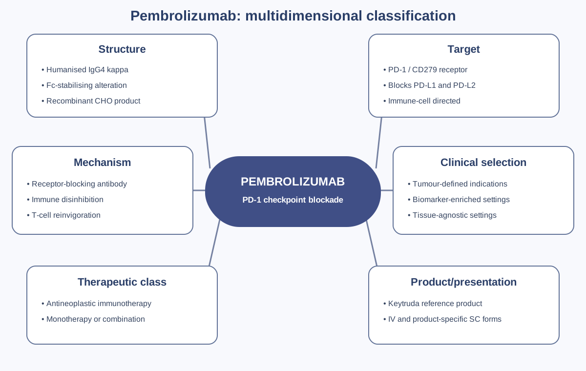
Figure 1. Pembrolizumab is a receptor-blocking, Fc-stabilised IgG4 immune-checkpoint antibody. Its classifications are simultaneous: molecular design limits unwanted effector function, PD-1 blockade disinhibits immunity, and indication-specific biomarkers determine patient selection.
Discovery and development history
PD-1 was identified in the early 1990s and later shown to function as an inhibitory immune receptor. Discovery of its ligands and the role of PD-1 signalling in peripheral tolerance provided a mechanistic explanation for how tumours suppress antigen-experienced T cells. Genetic and experimental work demonstrated that loss of this pathway can cause autoimmune phenotypes, foreshadowing the immune-mediated toxicities of therapeutic blockade.
Pembrolizumab originated as a humanised anti-PD-1 antibody programme known as MK-3475 and lambrolizumab. Early KEYNOTE-001 results showed durable tumour responses in advanced melanoma and later in non-small-cell lung cancer, with PD-L1 explored as an enrichment biomarker. The FDA granted accelerated approval for advanced melanoma in 2014. The EU authorised Keytruda on 17 July 2015.
Development expanded rapidly across melanoma, lung, head-and-neck, urothelial, renal, endometrial, cervical, gastric and other cancers, including perioperative and combination regimens. Certain authorisations use biomarkers such as PD-L1 expression, microsatellite instability or mismatch-repair deficiency; others are defined by histology, stage, treatment line or combination. “Pembrolizumab-eligible” is therefore not one universal biological category.
The programme also moved from intravenous treatment to an EU-authorised subcutaneous presentation containing berahyaluronidase alfa. This introduces presentation-specific dose, route and preparation controls. Current product information must be consulted because the indication portfolio and formulations continue to evolve.
PD-1 pathway biology
PD-1 is induced on activated T cells and is also expressed by other immune populations. After PD-L1 or PD-L2 binds PD-1, phosphorylated cytoplasmic motifs recruit phosphatases, particularly SHP-2, which attenuate proximal T-cell receptor and CD28 co-stimulatory signalling. The result can include reduced proliferation, cytokine production, metabolic fitness and cytotoxic activity.
In chronic antigen exposure, T cells can enter an exhausted state with altered transcriptional and epigenetic programmes. PD-1 blockade can expand and reinvigorate responsive progenitor-like exhausted T-cell populations, but does not simply return every cell to a naïve or fully functional state. Tumour antigenicity, antigen presentation, interferon signalling, immune-cell exclusion and suppressive myeloid or regulatory populations influence response.
Detailed mechanism of action
Pembrolizumab occupies PD-1 and sterically prevents binding by PD-L1 and PD-L2. By stopping inhibitory receptor engagement, it permits stronger signalling through the T-cell receptor and CD28 when antigen and co-stimulation are present. The antibody does not activate a T cell independently of antigen; it changes the threshold and persistence of an existing immune response.
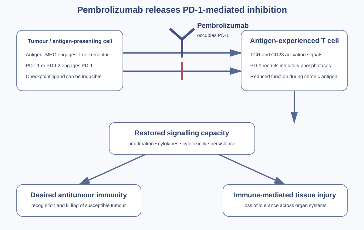
Figure 2. Tumour-cell antigen presentation and co-stimulation activate a T cell, while PD-L1/PD-L2 engagement of PD-1 recruits inhibitory phosphatase signalling. Pembrolizumab blocks the receptor–ligand interaction, restoring signalling capacity. The same loss of inhibitory control can injure normal tissues.
Antitumour consequence
Checkpoint release can increase T-cell proliferation, cytokine secretion, survival and cytotoxic killing. Tumour-cell death releases additional antigens, potentially broadening the immune response. Durable memory may persist after drug concentrations decline, helping explain long responses and why toxicity can continue after discontinuation.
Why IgG4 matters
PD-1 is expressed on the effector cells the therapy aims to preserve. An IgG1 Fc capable of strong complement activation or antibody-dependent cellular cytotoxicity could deplete PD-1-positive lymphocytes. The IgG4 framework reduces these functions. The stabilising Fc alteration limits IgG4 half-antibody exchange. Fc biology is reduced, not absent in every assay or tissue context.
Biomarkers and resistance
PD-L1 immunohistochemistry can enrich for response in specified settings, but assays, scoring algorithms, cut-offs and tumour types differ. Tumour proportion score and combined positive score are not interchangeable. Heterogeneity, sampling time and prior therapy affect results. MSI-high/dMMR tumours often generate many neoantigens and can respond across tissue types, but response is not guaranteed.
Primary or acquired resistance can involve absent antigenic targets, defective antigen presentation, beta-2-microglobulin loss, altered interferon signalling, T-cell exclusion, alternative checkpoints and immunosuppressive cells. These mechanisms differ in evidence and may coexist.
Immune-mediated adverse reactions
Immune-mediated adverse reactions can involve skin, bowel, liver, lung, endocrine organs, kidney, nervous system, heart, muscle, eye, blood or other tissues. They may occur during treatment or after it ends. Severity does not always correlate with time to onset, and routine laboratory changes can precede symptoms.
| Organ system | Important presentations | High-value evidence |
|---|---|---|
| Lung | Pneumonitis, organising pneumonia patterns | CT pattern, oxygenation, infection work-up, radiation and other pneumotoxic drugs |
| Gastrointestinal | Colitis, diarrhoea, perforation | Stool studies, endoscopy/biopsy, grade, hydration and complications |
| Liver | Immune-mediated hepatitis, cholangitic patterns | Serial enzymes/bilirubin, viral studies, imaging, biopsy and co-medication |
| Endocrine | Thyroiditis, hypo-/hyperthyroidism, hypophysitis, adrenal insufficiency, type 1 diabetes | Hormones, glucose/ketones, pituitary imaging and replacement requirement |
| Kidney | Interstitial nephritis and renal dysfunction | Creatinine trend, urine findings, biopsy and nephrotoxins |
| Neuromuscular/cardiac | Myositis, myasthenic syndrome, neuropathy, myocarditis | Troponin, CK, ECG, imaging, antibodies, respiratory measures and overlap |
| Skin | Rash, severe cutaneous reactions | Morphology, mucosa, body-surface area, biopsy and photographs |
Pneumonitis must be distinguished from infection, tumour progression, pulmonary embolism, heart failure and radiation injury. Colitis requires exclusion of infection and may worsen rapidly. Hepatitis can be hepatocellular or cholangitic and may respond differently to immunosuppression. Endocrine injury is often permanent and managed by hormone replacement rather than prolonged high-dose immunosuppression once acute inflammation is controlled.
Myocarditis is uncommon but can be rapidly fatal. Overlap with myositis and myasthenia-like syndromes is particularly important because weakness may precede cardiac or respiratory collapse. Normal early testing does not always exclude evolving disease.
Management is grade- and organ-specific and follows current product information and specialist guidance. It may include treatment interruption or permanent discontinuation, corticosteroids and other immunosuppression. Infliximab is not appropriate for every immune toxicity; for example, severe immune-mediated hepatitis requires different escalation. PV records should capture steroid dose, route, taper, second-line immunosuppression, prophylaxis and recurrence.
Special situations
Solid-organ transplant recipients can experience graft rejection when checkpoint inhibition disrupts transplant tolerance. Allogeneic haematopoietic-stem-cell transplantation before or after PD-1 blockade can be complicated by severe graft-versus-host disease and other immune effects. Pregnancy exposure raises concern because PD-1/PD-L1 contributes to maternal–fetal tolerance; current product-specific precautions apply.
Combination regimens create overlapping toxicity. Pembrolizumab plus chemotherapy can produce cytopenia, infection, hepatic or pulmonary events through several mechanisms. Axitinib and other kinase inhibitors can also cause hepatic, vascular and endocrine abnormalities. Attribution should support safe management rather than force a single suspect where interaction is plausible.
Pharmacovigilance architecture
The core safety record is a longitudinal immune phenotype: baseline disease and organ function, all anticancer and immune-modifying exposure, onset by organ, diagnostic exclusion, immunosuppression, recovery, taper, rechallenge and recurrence.
Cases should capture exact product/presentation, dose, route, dates and cycle; tumour and biomarkers; prior checkpoint inhibitors; radiation; transplant and autoimmune history; event grade and investigations; corticosteroid and second-line treatment; and outcome. Delayed events must be linked to prior pembrolizumab even after oncology follow-up has changed.
Signal analyses require stratification by tumour, regimen and surveillance intensity. A rise in thyroid testing can increase detection without changing biological risk. Combination therapy, longer survival and indication expansion alter exposed populations. Reporting counts are not incidence rates.
Medication errors include incorrect fixed versus weight-based regimens, schedule confusion, wrong presentation or route, subcutaneous/intravenous selection errors, and failure to recognise a prior immune toxicity before rechallenge. The long list of indications also creates biomarker and treatment-line selection errors.
Inspection and governance
An effective system should demonstrate current reference safety information; organ-specific follow-up; recognition of delayed and multi-organ toxicity; complete steroid and rechallenge data; linkage across oncology, emergency, endocrinology, cardiology, respiratory and primary care; and analysis by regimen and indication.
Common failures include coding “diarrhoea” without colitis work-up, treating hormone replacement as evidence that inflammation remains active, missing myocarditis–myositis overlap, closing a case when steroids start, and excluding pembrolizumab solely because onset followed discontinuation.
Practical checklist
- Verify tumour, stage, line, regimen and indication-specific biomarker.
- Record presentation, route, dose, schedule and every treatment date.
- Preserve baseline autoimmune, transplant, pulmonary, hepatic and endocrine status.
- Investigate new symptoms broadly; immune toxicity can affect any organ.
- Capture objective tests, infection exclusion, pathology and alternative causes.
- Document corticosteroid dose and taper, second-line immunosuppression and prophylaxis.
- Follow recovery, chronic sequelae, replacement therapy, rechallenge and recurrence.
- Maintain surveillance after the final dose.
Key takeaways
Pembrolizumab is an Fc-stabilised humanised IgG4 anti-PD-1 antibody. It blocks PD-1 interaction with PD-L1/PD-L2 and disinhibits antigen-dependent lymphocyte responses rather than directly killing tumour cells.
Response depends on tumour antigenicity, presentation, immune context and resistance biology. Biomarker method, scoring and indication are inseparable from interpretation.
Immune-mediated toxicity is multisystem, potentially delayed and sometimes permanent or fatal. Case quality depends on organ phenotype, exclusion of alternatives, immunosuppressive course and longitudinal outcome.
References
- European Medicines Agency. Keytruda: EPAR.
- European Medicines Agency. Keytruda EPAR product information. Current revision accessed September 2026.
- Ishida Y, Agata Y, Shibahara K, Honjo T. Induced expression of PD-1. EMBO J. 1992;11:3887–3895.
- Freeman GJ, Long AJ, Iwai Y, et al. Engagement of PD-1 by PD-L1 inhibits T-cell activation. J Exp Med. 2000;192:1027–1034.
- Hamid O, Robert C, Daud A, et al. Safety and tumor responses with lambrolizumab in melanoma. N Engl J Med. 2013;369:134–144.
- Garon EB, Rizvi NA, Hui R, et al. Pembrolizumab for non-small-cell lung cancer. N Engl J Med. 2015;372:2018–2028.
- Robert C, Schachter J, Long GV, et al. Pembrolizumab versus ipilimumab in advanced melanoma. N Engl J Med. 2015;372:2521–2532.
- Ribas A, Wolchok JD. Cancer immunotherapy using checkpoint blockade. Science. 2018;359:1350–1355.
- Pauken KE, Wherry EJ. Overcoming T cell exhaustion in infection and cancer. Trends Immunol. 2015;36:265–276.
- Postow MA, Sidlow R, Hellmann MD. Immune-related adverse events. N Engl J Med. 2018;378:158–168.
- Johnson DB, Balko JM, Compton ML, et al. Fulminant myocarditis with combined checkpoint blockade. N Engl J Med. 2016;375:1749–1755.
- Brahmer JR, Lacchetti C, Schneider BJ, et al. Management of immune-related adverse events. J Clin Oncol. 2018;36:1714–1768.
Regulatory Note
This is an educational scientific and pharmacovigilance review, not prescribing advice. Pembrolizumab indications, biomarker requirements, combinations, presentations, monitoring and toxicity-management instructions change over time and differ by jurisdiction. Consult current product-specific information and specialist guidance. Immune-mediated events may occur after discontinuation.