Ranibizumab: Classification, History, Mechanism of Action, Safety and Pharmacovigilance
- Ranibizumab: Classification, History, Mechanism of Action, Safety and Pharmacovigilance
- Multidimensional classification
- VEGF-A in retinal disease
- Why a Fab fragment was used
- Mechanism of action
- Development and regulatory evolution
- Intravitreal treatment and the ocular safety environment
- Injection-related adverse events
- Intraocular inflammation
- Intraocular pressure
- Ocular haemorrhage and common post-injection findings
- Systemic anti-VEGF considerations
- Immunogenicity
- Pregnancy and reproductive considerations
- Biosimilars and traceability
- Pharmacovigilance case assessment
- Signal detection and aggregate review
- Periodic benefit-risk evaluation
- Risk management and operational controls
- Potential failure modes
- Inspection and governance perspective
- Practical checklist
- Key Takeaways
- References
- Regulatory Note
Ranibizumab is an anti-vascular endothelial growth factor A (VEGF-A) antibody fragment developed specifically for intravitreal use. It is not a complete IgG molecule. It consists of the antigen-binding Fab portion of a humanised monoclonal antibody and therefore lacks an Fc region. This structural decision makes ranibizumab an instructive contrast with systemic full-length anti-VEGF antibodies: the target biology is related, but the product, route, distribution and dominant safety problems are different.
The pharmacovigilance profile is therefore best understood by separating drug effect from injection-procedure effect. VEGF inhibition explains the desired reduction in vascular leakage and pathological neovascularisation. By contrast, endophthalmitis, retinal tear, retinal detachment and traumatic cataract are linked primarily to intravitreal administration. Intraocular pressure elevation and intraocular inflammation sit between these categories because they can be influenced by the procedure, repeated treatment and patient-specific ocular anatomy.
Multidimensional classification
| Classification axis | Ranibizumab classification | Scientific or PV significance |
|---|---|---|
| Molecular class | Humanised monoclonal-antibody Fab fragment | Lacks Fc region and is substantially smaller than a full IgG antibody |
| Target | VEGF-A | Neutralises a key mediator of angiogenesis and vascular permeability |
| Functional class | Anti-angiogenic / vascular-permeability inhibitor | Reduces pathological vessel growth and leakage in retinal disease |
| Route | Intravitreal injection | Dominant safety burden includes ocular procedure-related events |
| Distribution strategy | Local ocular delivery with limited systemic exposure | Ocular efficacy is achieved without systemic antineoplastic dosing |
| Therapeutic class | Ophthalmological anti-VEGF biological | Used across several retinal vascular and neovascular conditions |
| Product category | Biological medicinal product with authorised biosimilars | Exact product and batch are required for biological traceability |
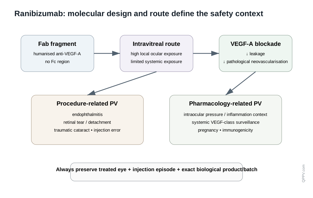
Figure 1. Ranibizumab combines a VEGF-A-neutralising Fab fragment with intravitreal delivery. The absence of Fc and the local ocular route distinguish its pharmacology and safety context from full-length systemic anti-VEGF antibodies.
VEGF-A in retinal disease
VEGF-A is a signalling protein that promotes endothelial-cell survival, vascular permeability and angiogenesis. These functions are essential during development and tissue repair but become pathological when VEGF is produced inappropriately or excessively within the eye.
The retina is metabolically demanding and highly sensitive to disruption of oxygen delivery. Ischaemia, inflammation and age-related tissue changes can increase VEGF expression. VEGF then increases permeability of retinal vessels and can promote formation of fragile abnormal vessels. Fluid accumulation, haemorrhage and disruption of retinal architecture impair photoreceptor function and therefore vision.
Neovascular age-related macular degeneration
In neovascular or “wet” age-related macular degeneration, abnormal choroidal vessels grow beneath or into the macular region. These vessels are structurally abnormal and leak fluid or blood. VEGF-A blockade reduces vascular permeability and neovascular activity, allowing retinal fluid to resolve and limiting further structural damage.
Diabetic macular oedema
Diabetic retinal microvascular injury increases permeability and disrupts the blood-retinal barrier. VEGF contributes to leakage and macular thickening. Anti-VEGF therapy therefore targets a major mediator of oedema rather than the systemic metabolic cause of diabetes itself.
Retinal vein occlusion
Venous obstruction causes retinal congestion and ischaemia, increasing VEGF production and vascular leakage. Macular oedema can then impair vision. VEGF blockade reduces leakage but does not remove the underlying vascular occlusion.
Choroidal neovascularisation from other causes
VEGF-driven choroidal neovascularisation can occur in pathological myopia and other disorders. The common pharmacological target is abnormal VEGF-dependent vessel growth, but the underlying disease and expected natural history differ.
Why a Fab fragment was used
A complete IgG antibody contains two antigen-binding arms and an Fc region. Ranibizumab retains one antigen-binding Fab fragment but not the Fc. It is therefore smaller and monovalent.
The Fab was affinity matured to bind VEGF-A strongly and neutralise biologically active isoforms. Its smaller size was intended to support retinal penetration after intravitreal injection. The absence of Fc also removes Fc-receptor and complement effector functions that are unnecessary for soluble VEGF neutralisation.
This is an important conceptual distinction. Ranibizumab is not designed to coat a cell and recruit immune destruction. Its role is ligand sequestration: bind soluble VEGF-A so that VEGF receptors on endothelial cells cannot be activated.
Mechanism of action
VEGF-A normally binds VEGF receptors, especially VEGFR-1 and VEGFR-2, on endothelial cells. Receptor activation promotes endothelial proliferation, survival, permeability and migration. Ranibizumab binds VEGF-A before it reaches these receptors.
The pharmacological sequence is therefore:
- pathological retinal or choroidal tissue produces VEGF-A;
- VEGF-A would normally activate endothelial VEGF receptors;
- ranibizumab binds VEGF-A within the ocular compartment;
- receptor activation falls;
- vascular leakage and neovascular signalling decrease;
- retinal fluid can resolve and visual function may stabilise or improve if irreversible structural damage has not already occurred.
The medicine does not regenerate lost photoreceptors or reverse established fibrosis. This distinction is important when evaluating apparent lack of efficacy in advanced disease.
Development and regulatory evolution
Ranibizumab was developed from an anti-VEGF antibody lineage but optimised as an affinity-matured Fab for ophthalmic use. Early pivotal studies in neovascular AMD demonstrated that repeated intravitreal VEGF inhibition could do more than slow visual loss: many treated patients maintained vision, and a meaningful proportion improved.
The European Union authorised ranibizumab in 2007. Its indication set subsequently expanded from neovascular AMD to diabetic macular oedema, macular oedema after retinal vein occlusion and choroidal neovascularisation from additional causes. Current EMA product information, updated in September 2026, should be used for the exact authorised disease, dosing strategy and monitoring requirements.
The active substance is now also available through authorised biosimilars. This changes product-traceability requirements without changing the fundamental scientific mechanism.
Intravitreal treatment and the ocular safety environment
Intravitreal administration places a small volume of medicine directly into the vitreous cavity. This achieves high local exposure but creates a procedural risk that is not shared by orally or intravenously administered medicines. The safety system must therefore reconstruct not only the molecule and dose, but also the injection episode.
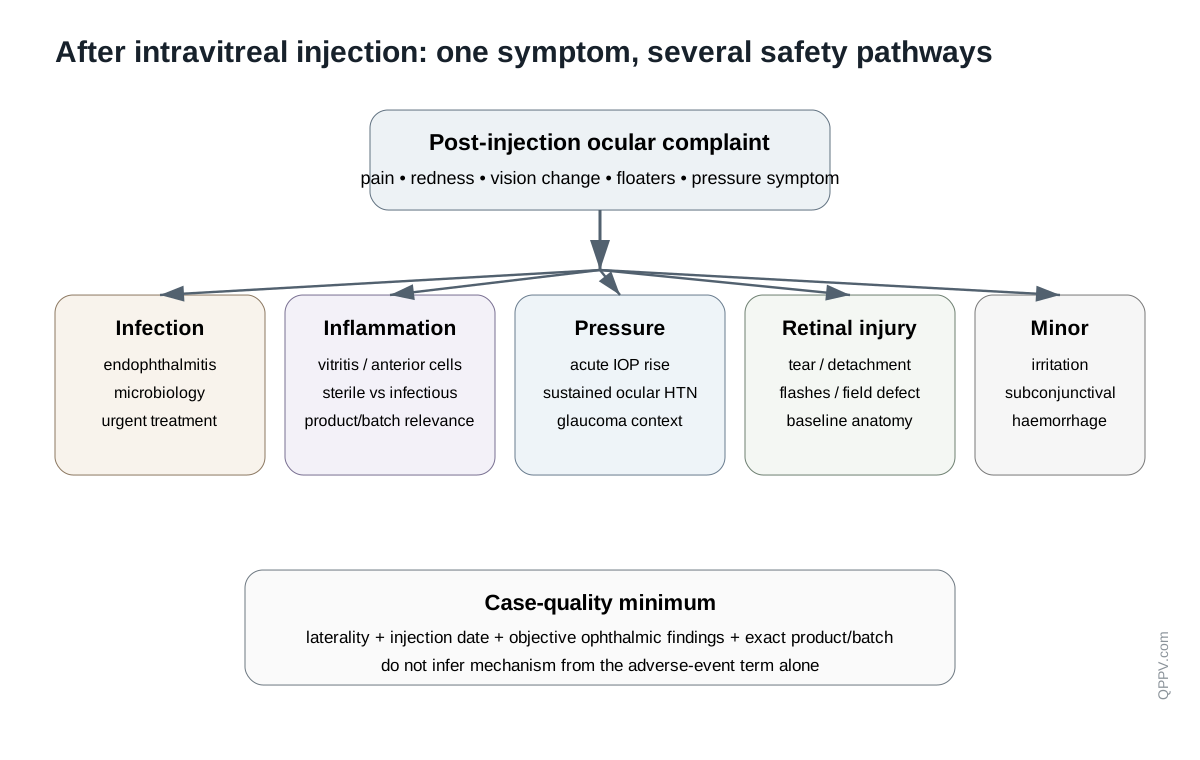
Figure 2. Ranibizumab PV separates pharmacological anti-VEGF effects from complications of intravitreal injection. The same post-injection symptom can require assessment for infection, inflammation, pressure change, retinal injury or expected transient discomfort.
Injection-related adverse events
Endophthalmitis
Endophthalmitis is an intraocular infection and is one of the most important serious complications of intravitreal injection. It can present with eye pain, worsening redness, photophobia and reduced vision, but symptom patterns vary. Delay in recognition can result in permanent visual loss.
Case follow-up should collect injection date and eye, onset, aseptic procedure details where available, symptoms, visual acuity, slit-lamp/vitreous findings, microbiology, intravitreal antimicrobial treatment, surgery and final visual outcome. The distinction between infectious endophthalmitis and sterile intraocular inflammation is clinically important and should not be inferred from the adverse-event term alone.
Retinal tear and retinal detachment
Mechanical entry into the eye can be associated with retinal tear or rhegmatogenous retinal detachment. Symptoms such as flashes, new floaters or a curtain-like field defect require urgent ophthalmic assessment. PV reports should capture retinal examination, imaging, previous retinal disease, high myopia, prior surgery and treatment outcome.
A retinal detachment developing after injection is temporally associated with a procedure but may also arise from strong pre-existing anatomical risk factors. Causality should therefore preserve both the injection relationship and the patient's ocular background.
Iatrogenic traumatic cataract
Accidental lens injury during intravitreal injection can produce traumatic cataract. This is a procedural event rather than a VEGF-A pharmacodynamic effect. Case narratives should therefore retain needle/procedure details, lens findings, timing and subsequent cataract management.
Intraocular inflammation
Inflammatory events include anterior chamber inflammation, vitritis and broader intraocular inflammatory syndromes. The key differential is infectious versus non-infectious inflammation.
High-value information includes pain, redness, photophobia, visual change, anterior chamber cells/flare, vitreous cells, hypopyon, microbiology, corticosteroid or antimicrobial treatment and response. Product/batch information is particularly important when multiple sterile inflammatory cases appear clustered.
Intraocular pressure
Transient rises in intraocular pressure can occur immediately after intravitreal injection because fluid is added to a closed ocular compartment. Sustained pressure elevation has also been observed during repeated intravitreal anti-VEGF treatment.
The acute and chronic phenomena should be separated. An immediate post-injection pressure rise should be interpreted alongside injection volume, optic-nerve perfusion and glaucoma history. Sustained elevation requires longitudinal pressure data, cumulative injection history and glaucoma therapy.
Current EMA product information specifically identifies both transient and sustained IOP increases and requires appropriate monitoring and management. For PV, the important variables are baseline pressure, post-injection measurements, optic-nerve status, glaucoma history and whether intervention was needed.
Ocular haemorrhage and common post-injection findings
Conjunctival haemorrhage, retinal haemorrhage, vitreous floaters, eye irritation and transient discomfort are common in populations receiving intravitreal therapy. These events vary greatly in clinical importance. A small conjunctival haemorrhage after needle entry is not equivalent to sight-threatening intraocular bleeding.
Aggregate analyses should therefore avoid pooling all “haemorrhage” terms without anatomical and severity stratification.
Systemic anti-VEGF considerations
Ranibizumab is intended for local ocular delivery, but a fraction reaches the systemic circulation. Systemic exposure is substantially lower than with intravenous anti-VEGF therapy and the Fab is cleared relatively rapidly, yet systemic VEGF suppression remains a theoretical and monitored safety consideration.
Arterial thromboembolic events
VEGF contributes to vascular homeostasis, so arterial thromboembolic events such as stroke and myocardial infarction have long been monitored across intravitreal VEGF inhibitors. Current product information discusses a potential systemic class risk rather than treating every vascular event as established ranibizumab causality.
PV assessment should capture age, prior stroke or myocardial infarction, diabetes, hypertension, smoking, bilateral treatment, timing from injection and other vascular risk factors. The treated population itself often has substantial baseline cardiovascular risk.
Bilateral treatment
Same-day bilateral treatment increases the amount of drug administered in one clinical encounter and creates two independent ocular procedure sites. EMA information notes limited data that do not suggest increased systemic adverse events compared with unilateral treatment, but PV should still record whether one or both eyes were treated because this affects exposure and procedural interpretation.
A bilateral endophthalmitis or inflammation scenario has especially high product-quality and procedural significance and should prompt detailed batch, preparation and aseptic-technique investigation.
Immunogenicity
As a biological protein, ranibizumab can generate anti-drug immune responses. The clinical relevance of detected antibodies is not always straightforward because assay sensitivity, ocular inflammation and systemic exposure influence interpretation.
Suspected immunogenicity cases should therefore not be based on an antibody test alone. Relevant clinical evidence includes recurrent intraocular inflammation, loss of response, hypersensitivity, product switch, timing and assay details.
Pregnancy and reproductive considerations
Although systemic exposure after intravitreal treatment is low, VEGF has essential physiological roles in angiogenesis and embryofetal development. Pregnancy exposure should therefore be documented carefully, including gestational timing, treated eye(s), number of injections, maternal retinal disease, co-exposures and outcome.
The low systemic dose does not justify assuming zero fetal exposure, while the theoretical VEGF-development concern does not establish that an adverse pregnancy outcome was caused by treatment. Current regional product information should guide clinical recommendations.
Biosimilars and traceability
Authorised ranibizumab biosimilars are expected to have no clinically meaningful differences from the reference medicine in authorised uses after regulatory biosimilarity assessment. Pharmacovigilance nevertheless requires exact brand/product and batch capture for biological traceability.
This is particularly important in ophthalmology because an apparent cluster of sterile inflammation, endophthalmitis or device-related complaints may be product- or batch-specific. Coding every exposure only as “ranibizumab” can destroy the information needed to investigate such a cluster.
Pharmacovigilance case assessment
Ranibizumab case assessment should start by identifying the treated eye, indication and exact injection episode. Ocular medicine safety often fails when the database records only the active substance and adverse-event term while losing laterality, procedure timing, ocular history and product batch.
Event-specific follow-up priorities
| Event | High-value follow-up information |
|---|---|
| Endophthalmitis | Treated eye, injection date, onset, pain/redness/vision loss, exam, microbiology, intravitreal antibiotics, surgery, final visual outcome |
| Intraocular inflammation | Anterior/vitreous findings, hypopyon, microbiology, steroid/antibiotic treatment, recurrence, product/batch |
| Retinal tear/detachment | Laterality, symptoms, retinal findings, high myopia, prior surgery, procedure timing, repair and outcome |
| Traumatic cataract | Injection details, lens injury evidence, timing, surgery and visual outcome |
| IOP increase | Baseline/post-dose pressure, glaucoma history, cumulative injections, optic-nerve status and treatment |
| Arterial thromboembolic event | Stroke/MI phenotype, vascular risk factors, timing, bilateral treatment, competing causes |
| Lack of efficacy | Indication, OCT/fluid trajectory, visual acuity, lesion activity, adherence to treatment schedule, irreversible structural disease |
| Product-quality event | Exact product/biosimilar, batch, storage, syringe/device, preparation and aseptic procedure details |
Signal detection and aggregate review
Signal detection should distinguish events caused primarily by pharmacology from those caused primarily by the injection procedure. Endophthalmitis, retinal tear and traumatic cataract should not be interpreted as if they arise from VEGF neutralisation. Conversely, a vascular-event analysis should not be mixed with local needle complications merely because both occurred after an injection.
Laterality is an important analytical variable. Recurrent inflammation in the same treated eye, bilateral same-day events and events occurring in an untreated fellow eye have different causal implications.
Product and batch stratification is also critical for biological and sterile-inflammation signals. A cluster can disappear statistically if multiple ranibizumab products are pooled under the same INN.
Periodic benefit-risk evaluation
Periodic evaluation should connect exposure with indication, injection frequency, bilateral treatment and product identity. Important domains include endophthalmitis, intraocular inflammation, retinal tear/detachment, traumatic cataract, IOP changes, ocular haemorrhage, arterial thromboembolic events, immunogenicity, pregnancy exposure, medication/procedure errors and product-quality complaints.
The benefit side must be disease-specific. Maintaining vision in neovascular AMD, reducing diabetic macular oedema and restoring retinal anatomy after vein-occlusion-related oedema are related but distinct treatment objectives. Apparent loss of efficacy should therefore be interpreted against expected natural history and structural retinal damage for the specific disease.
Risk management and operational controls
Current regional product information governs aseptic technique, injection intervals, ocular monitoring and management of complications. Pharmacovigilance operations should support those requirements while distinguishing mandated product-information instructions from locally recommended clinical practice.
Useful controls include mandatory laterality fields, structured capture of injection date and treated eye, targeted endophthalmitis/inflammation follow-up, baseline and post-injection IOP fields, exact biosimilar/reference product identification and batch capture. Device or syringe complaints should be reconciled with product-quality systems.
Potential failure modes
The following are illustrative scenarios rather than published inspection findings:
- Endophthalmitis is reported without identifying which eye was injected.
- Sterile intraocular inflammation is classified as infection despite negative microbiology and steroid-responsive course.
- A retinal detachment is treated as a pharmacological anti-VEGF effect without preserving the injection-procedure relationship and baseline retinal risk.
- A pressure signal combines immediate transient post-injection rises with chronic sustained ocular hypertension.
- A stroke case is attributed to ranibizumab without documenting the patient's substantial baseline vascular risk.
- Apparent lack of efficacy is assessed without OCT findings or recognition of irreversible macular fibrosis.
- A cluster of inflammation cannot be investigated because only the INN, not product and batch, was recorded.
Inspection and governance perspective
An inspector assessing ranibizumab pharmacovigilance would be interested in whether the system preserves the procedural information needed to evaluate an intravitreal biological. Evidence may include laterality conventions, targeted ocular follow-up forms, product/batch traceability, quality-complaint reconciliation, signal analyses separating infection from sterile inflammation, and methodology for procedure-related versus systemic events.
Effectiveness is demonstrated when serious ocular cases are clinically reconstructable. A procedure stating “record administration details” is not effective if endophthalmitis cases lack treated eye, injection date or product batch.
Practical checklist
For a ranibizumab case or aggregate review, confirm:
- exact indication and treated eye;
- injection date, dose and unilateral versus bilateral treatment;
- exact product, biosimilar/reference status and batch;
- baseline ocular history including glaucoma, high myopia and prior surgery;
- objective ophthalmic findings and visual-acuity outcome;
- infectious versus sterile inflammatory evidence;
- baseline and post-injection IOP for pressure events;
- cardiovascular risk profile for systemic vascular events;
- OCT or imaging evidence for apparent lack of efficacy;
- device, storage, preparation and aseptic-procedure details for quality/procedure events.
Key Takeaways
Ranibizumab is a humanised anti-VEGF-A Fab fragment designed for intravitreal use. Its lack of Fc, smaller size and local ocular route distinguish it from full-length systemic anti-VEGF antibodies even though the soluble ligand target is shared.
Its pharmacovigilance profile is dominated by the interface between drug and procedure. Endophthalmitis, retinal tear or detachment, traumatic cataract, intraocular inflammation and pressure changes require detailed injection-context assessment, while systemic arterial thromboembolic risk remains a monitored class consideration against a high-risk background population. Exact product and batch traceability is increasingly important as biosimilars expand.
References
- European Medicines Agency. Ranibizumab: EPAR and current product information. Product information last updated 8 September 2026. https://www.ema.europa.eu/en/medicines/human/EPAR/lucentis
- European Medicines Agency. Ranibizumab product information. Current SmPC includes biological traceability, intravitreal injection-related reactions and intraocular-pressure precautions. https://www.ema.europa.eu/en/documents/product-information/lucentis-epar-product-information_en.pdf
- Rosenfeld PJ, Brown DM, Heier JS, et al. Ranibizumab for neovascular age-related macular degeneration. N Engl J Med. 2006;355:1419-1431. doi:10.1056/NEJMoa054481.
- Brown DM, Kaiser PK, Michels M, et al. Ranibizumab versus verteporfin for neovascular age-related macular degeneration. N Engl J Med. 2006;355:1432-1444. doi:10.1056/NEJMoa062655.
- European Medicines Agency. Ranivisio EPAR. Biosimilar ranibizumab regulatory overview and comparability conclusions. https://www.ema.europa.eu/en/medicines/human/EPAR/ranivisio
Regulatory Note
Authorised indications, dosing intervals, injection procedures, contraindications, warnings and monitoring requirements vary by jurisdiction and may change. This article does not replace current regional product information. Regulatory statements were checked against current EMA information available in September 2026. Operational recommendations are pharmacovigilance practice unless explicitly identified as regulatory requirements.