Ranibizumab Port-Delivery Implant: Device, Safety and Pharmacovigilance
- Ranibizumab Port-Delivery Implant: Device, Safety and Pharmacovigilance
- Product-device architecture
- Why continuous delivery changes the PV model
- Current U.S. treatment context
- Endophthalmitis as the defining serious risk
- Implant- and procedure-related safety domains
- The refill-exchange procedure as a repeated exposure
- Drug-related versus device-related causality
- Pharmacovigilance case assessment
- Signal detection: link drug and device datasets
- Medication and device-use errors
- Illustrative failure modes
- Inspection and governance perspective
- Key Takeaways
- References
- Regulatory Note
A ranibizumab port-delivery system converts intermittent intravitreal dosing into continuous ocular delivery from an implanted reservoir. The active molecule remains ranibizumab, an anti-VEGF-A Fab fragment, but the safety architecture is fundamentally different from standard intravitreal injection. The patient undergoes implant surgery, lives with a permanent ocular device and later undergoes repeated refill-exchange procedures.
Pharmacovigilance must therefore consider three interacting sources of harm: drug pharmacology, implant/device behaviour, and procedure-related complications. A report of vision loss cannot be assessed credibly until those layers are separated.
Product-device architecture
| Layer | Function | PV significance |
|---|---|---|
| Ranibizumab | Neutralises VEGF-A | Controls retinal vascular leakage and neovascular signalling |
| Ocular implant | Stores and continuously releases ranibizumab | Creates long-duration exposure and permanent device-related risks |
| Surgical implantation | Places implant through ocular tissues | Introduces perioperative infection, haemorrhage and retinal risks |
| Refill-exchange procedure | Replaces reservoir contents periodically | Creates repeated procedural and device-access risk |
| Refill needle / handling system | Enables controlled exchange of implant contents | Device malfunction, misalignment or technique errors can affect safety |
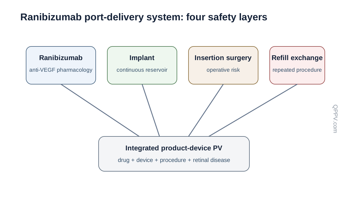
Figure 1. Safety arises from the active drug, implanted reservoir, insertion surgery and repeated refill-exchange procedures. These layers must remain separately traceable.
Why continuous delivery changes the PV model
Standard intravitreal ranibizumab creates a repeated sequence of injection exposures. The port-delivery system creates a persistent device state between procedures. This changes chronology: an event may occur days after surgery, months into implant residence, immediately after refill, or after cumulative device manipulation.
The correct temporal question is therefore not simply “how long after the last ranibizumab dose?” It is “where was the patient in the implant lifecycle when the event occurred?”
Current U.S. treatment context
Current U.S. labelling includes patients with neovascular age-related macular degeneration, diabetic macular oedema and diabetic retinopathy who have previously responded to at least two intravitreal VEGF-inhibitor injections. Refill intervals differ by indication, with approximately six-month refill-exchange intervals for wet AMD and DME and approximately nine-month intervals for diabetic retinopathy under current labelling.
These distinctions matter for PV because procedure frequency changes cumulative procedural exposure. A patient undergoing more frequent refill-exchange procedures has a different exposure history from a patient with a longer refill interval.
Endophthalmitis as the defining serious risk
Endophthalmitis is a severe intraocular infection that can threaten vision. Current U.S. labelling carries a boxed warning and states that the implant has been associated with a higher rate of endophthalmitis than monthly intravitreal ranibizumab injections.
A suspected case requires urgent reconstruction of surgery or refill timing, conjunctival status, symptoms, microbiology, treatment, visual outcome and whether implant removal or other surgery occurred. Device and procedure details are not optional background—they are part of the causal evidence.
Implant- and procedure-related safety domains
Rhegmatogenous retinal detachment
Retinal detachment can occur in relation to ocular surgery or subsequent device/procedure complications. A case should preserve the retinal break or detachment pattern, timing from implantation or refill, surgical findings, management and visual outcome. Without those data, the event cannot be separated from progression of underlying retinal disease or unrelated retinal pathology.
Vitreous haemorrhage
Vitreous haemorrhage may occur around implantation or other ocular procedures and can also arise from the underlying retinal vascular disease. Assessment should capture antithrombotic therapy, operative timing, retinal neovascular activity, need for vitrectomy and whether the haemorrhage followed insertion, refill or another intervention.
Implant dislocation and septum complications
A permanent ocular implant creates mechanical failure modes that do not exist with standalone intravitreal injections. Implant dislocation, septum dislodgement and observed device damage require explicit device-event coding and should trigger collection of procedure details, device position, refill technique, imaging and corrective surgery.
A device complaint with no immediate clinical consequence is still relevant because repeated reports can identify a quality or use-related signal before serious injury becomes common.
Conjunctival erosion, retraction and bleb formation
The implant traverses ocular tissues and depends on durable conjunctival coverage. Erosion or retraction can expose the implant and may increase infection risk. Safety assessment should therefore connect conjunctival findings with later endophthalmitis rather than treating them as unrelated events.
Intraocular inflammation
Intraocular inflammation can occur after implantation and may present with cells, flare, pain, photophobia or reduced vision. Current U.S. safety information was strengthened in 2025 to highlight postoperative intraocular inflammation, particularly in the early period after implantation. Differential diagnosis must include infection because sterile inflammation and infectious endophthalmitis can overlap clinically.
Postoperative visual decline
A transient postoperative decrease in visual acuity can occur after implant surgery, but persistent or severe vision loss requires evaluation for haemorrhage, retinal detachment, infection, inflammation, device position and other ocular causes. “Expected postoperative change” should never become a blanket explanation for an incompletely investigated serious event.
The refill-exchange procedure as a repeated exposure
The refill-exchange procedure is not equivalent to a routine injection. A dedicated refill needle exchanges the reservoir contents while interacting mechanically with the implant septum. Technique, alignment, sterility, device integrity and residual fluid handling all matter.
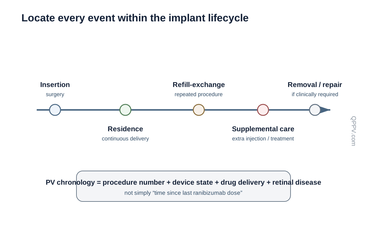
Figure 2. Safety assessment should locate each event within the implant lifecycle: insertion, residence, refill-exchange, supplemental treatment, complication management or removal.
Repeated procedures create cumulative opportunity for error. PV should therefore record the procedure number, date, interval from prior refill, operator-reported difficulty, device findings and whether the event followed an uncomplicated or abnormal refill.
Drug-related versus device-related causality
Ranibizumab pharmacology can contribute to ocular and systemic anti-VEGF considerations, but many defining risks of the port-delivery system are procedural or mechanical. A serious case may involve both layers. For example, reduced vision after refill could reflect inflammation, infection, haemorrhage, retinal disease activity or insufficient drug delivery due to a device problem.
The appropriate causality model is therefore drug + device + procedure + disease, not a forced choice between “adverse drug reaction” and “device event.”
Pharmacovigilance case assessment
The case should be reconstructed around the implant lifecycle rather than only the last drug exposure.
| Event | High-value follow-up |
|---|---|
| Endophthalmitis | Timing from implantation/refill, conjunctival status, symptoms, cultures/PCR, intravitreal/systemic treatment, surgery, visual outcome |
| Retinal detachment | Retinal break, imaging/exam findings, timing from procedure, repair, visual outcome |
| Vitreous haemorrhage | Procedure timing, antithrombotics, retinal disease activity, vitrectomy, recovery |
| Implant dislocation/damage | Device position, imaging, procedure details, refill difficulty, corrective surgery |
| Septum dislodgement | Refill number, needle alignment, device findings, leakage/delivery concerns, intervention |
| Conjunctival erosion/retraction | Implant exposure, infection signs, repair, later endophthalmitis |
| Intraocular inflammation | Implantation/refill timing, anterior/vitreous findings, infection exclusion, corticosteroid treatment, outcome |
| Loss of efficacy | Disease activity, refill timing, supplemental injections, device integrity, imaging, adherence to refill schedule |
Signal detection: link drug and device datasets
A product-device safety programme should not split related evidence into isolated databases without reconciliation. Endophthalmitis reports may begin as adverse-event cases, device complaints or surgical reports. Septum or refill-needle problems may appear in device surveillance before a clinical event is recognised. Signal detection therefore requires linkage between pharmacovigilance, product quality, device vigilance and medical-information sources.
Useful aggregate dimensions include implant age, indication, refill number, time since last procedure, operator-reported refill difficulty, conjunctival complications and device replacement/removal status.
Medication and device-use errors
Potential failure modes include using an incorrect refill technique, incomplete exchange of implant contents, air introduction, misalignment of the refill needle, delayed refill, wrong ranibizumab presentation, and documentation that fails to distinguish supplemental intravitreal injection from implant refill.
These events should be assessed for clinical consequence even when no adverse reaction is initially apparent.
Illustrative failure modes
The following are hypothetical examples, not published inspection findings:
- An endophthalmitis case is processed as a generic ranibizumab infection event without recording whether it followed implantation or refill.
- A device complaint involving refill resistance is closed because the patient is asymptomatic, preventing trend detection.
- Vision loss is labelled “disease progression” despite contemporaneous implant displacement.
- A supplemental intravitreal ranibizumab injection is mistaken for a routine implant refill in the exposure chronology.
- Conjunctival erosion and later endophthalmitis are coded in separate cases without linkage.
Inspection and governance perspective
An inspector or quality reviewer would be expected to examine whether adverse-event, device-complaint and product-quality processes communicate effectively. Evidence could include reconciliation procedures, device identifiers, procedure-specific follow-up forms, complaint trending, signal assessments, corrective-action records and documentation of escalation when device integrity may affect drug delivery or infection risk.
The central governance question is whether the organisation can reconstruct the complete drug-device-procedure trajectory for a patient.
Key Takeaways
The ranibizumab port-delivery implant is a combined medicinal-product and device safety problem. Ranibizumab provides continuous anti-VEGF activity, while the implant and repeated procedures introduce risks that standard intravitreal injections do not share to the same degree.
Endophthalmitis, retinal detachment, vitreous haemorrhage, conjunctival erosion/retraction, implant dislocation, septum complications, intraocular inflammation and refill-related errors all require device- and procedure-specific follow-up. Pharmacovigilance is strongest when drug, device, quality and surgical evidence are integrated rather than reviewed in isolation.
References
- U.S. Food and Drug Administration. Ranibizumab injection for intravitreal use via ocular implant: current prescribing information. Current SPL/label information, 2025–2026.
- U.S. Food and Drug Administration. SUSVIMO (ranibizumab injection) label. Indications include neovascular AMD, diabetic macular oedema and diabetic retinopathy in patients previously responsive to intravitreal VEGF inhibition.
- Campochiaro PA, et al. Ranibizumab port delivery system for neovascular age-related macular degeneration: pivotal clinical development literature.
- Khanani AM, et al. Long-term evaluation of continuous ranibizumab delivery through the port-delivery system in neovascular AMD.
Regulatory Note
This article describes the U.S.-authorised ranibizumab port-delivery implant. Device availability, indications, refill intervals, warnings and procedural instructions are jurisdiction-specific and may change. Current U.S. information was checked in September 2026. Operational recommendations are pharmacovigilance and quality practice unless explicitly identified as regulatory requirements.