Rituximab: Classification, History, Mechanism of Action and Pharmacovigilance
- Rituximab: Classification, History, Mechanism of Action and Pharmacovigilance
- Multidimensional classification
- Why CD20 became a therapeutic target
- Discovery and development history
- CD20 biology
- Detailed mechanism of action
- Clinical-use architecture
- Pharmacokinetics and pharmacodynamics
- Safety profile as a consequence of mechanism and context
- A pharmacovigilance model for rituximab
- Risk minimisation and evidence of effectiveness
- Important pharmacovigilance topics
- Regulatory and safety evolution
- Causality assessment by event type
- Inspection and governance perspective
- Practical checklist
- Key takeaways
- References
- Regulatory Note
Rituximab is one of the defining medicines of modern antibody therapeutics. It was the first monoclonal antibody approved in the United States for the treatment of cancer and helped establish that a lineage-associated cell-surface antigen could be used to eliminate a malignant cell population while preserving the capacity for later immune reconstitution. Its importance extends beyond oncology. By depleting CD20-positive B cells, rituximab also altered the treatment of antibody-mediated and B-cell-dependent inflammatory disease.
The apparent simplicity of the therapeutic concept—bind CD20 and remove B cells—conceals substantial biological and pharmacovigilance complexity. Rituximab does not kill cells through one invariant pathway. Complement, Fc-receptor-bearing effector cells, phagocytes and direct signalling can each contribute, and their relative importance varies with the target cell, tissue compartment, disease, tumour burden, host immune function, concomitant treatment and exposure. The same biology that produces benefit can cause infusion-related reactions, infection, viral reactivation, impaired vaccine responses, cytopenias and prolonged secondary immune deficiency.
This article treats rituximab as a product, a molecular intervention and a longitudinal safety-management problem. It focuses on the European reference product MabThera while recognising that rituximab is available through multiple brands and biosimilars. Product-specific prescribing information must therefore remain the immediate source for authorised indications, formulations, administration and risk-minimisation requirements.
Multidimensional classification
No single label adequately classifies rituximab. “Monoclonal antibody,” “anti-CD20 therapy,” “B-cell-depleting agent” and “antineoplastic medicine” are all correct, but each describes a different axis.
| Classification axis | Rituximab classification | Scientific or PV significance |
|---|---|---|
| Molecular class | Monoclonal antibody; immunoglobulin G1 kappa | IgG1 Fc supports complement activation and engagement of Fc gamma receptors |
| Species composition | Chimeric mouse–human antibody | Murine variable regions provide antigen recognition; human constant regions support human effector functions; anti-drug antibodies remain possible |
| Glycosylation | Glycosylated protein | Fc glycan characteristics can influence effector function and are critical quality attributes |
| Production platform | Recombinant expression in Chinese hamster ovary cells | Manufacturing consistency, process controls and comparability are integral to product quality |
| Antibody-generation class | First-generation, unmodified anti-CD20 antibody | Distinct from later humanised, fully human or Fc-engineered anti-CD20 antibodies |
| CD20 functional class | Type I anti-CD20 antibody | Characteristically promotes CD20 redistribution into membrane microdomains and strong complement engagement |
| Target | CD20, a B-cell surface protein encoded by MS4A1 | Targets most mature B-cell stages but generally spares haematopoietic stem cells and terminally differentiated plasma cells |
| Proximal pharmacology | Target-cell opsonisation and CD20 organisation | Creates a platform for complement and Fc-dependent effector mechanisms |
| Effector mechanisms | Complement-dependent cytotoxicity; antibody-dependent cellular cytotoxicity; antibody-dependent cellular phagocytosis; direct signalling/cell-death effects | Contributions are context-dependent and should not be represented as a single proven dominant mechanism in all patients |
| Functional therapeutic class | B-cell-depleting therapy | Explains both antitumour activity and immunomodulatory effects |
| Therapeutic/regulatory class | Primarily antineoplastic in ATC classification; also used in authorised inflammatory/autoimmune indications | Indication changes baseline risk, co-therapy, dosing, monitoring and risk communication |
| Regulatory product category | Biological medicinal product; reference product and active substance for authorised biosimilars | Brand, formulation and route must be traceable in safety reports |
| Formulation/route | Intravenous formulations; a product-specific subcutaneous formulation for certain haematological indications | Route and strength differences create administration and medication-error risks |
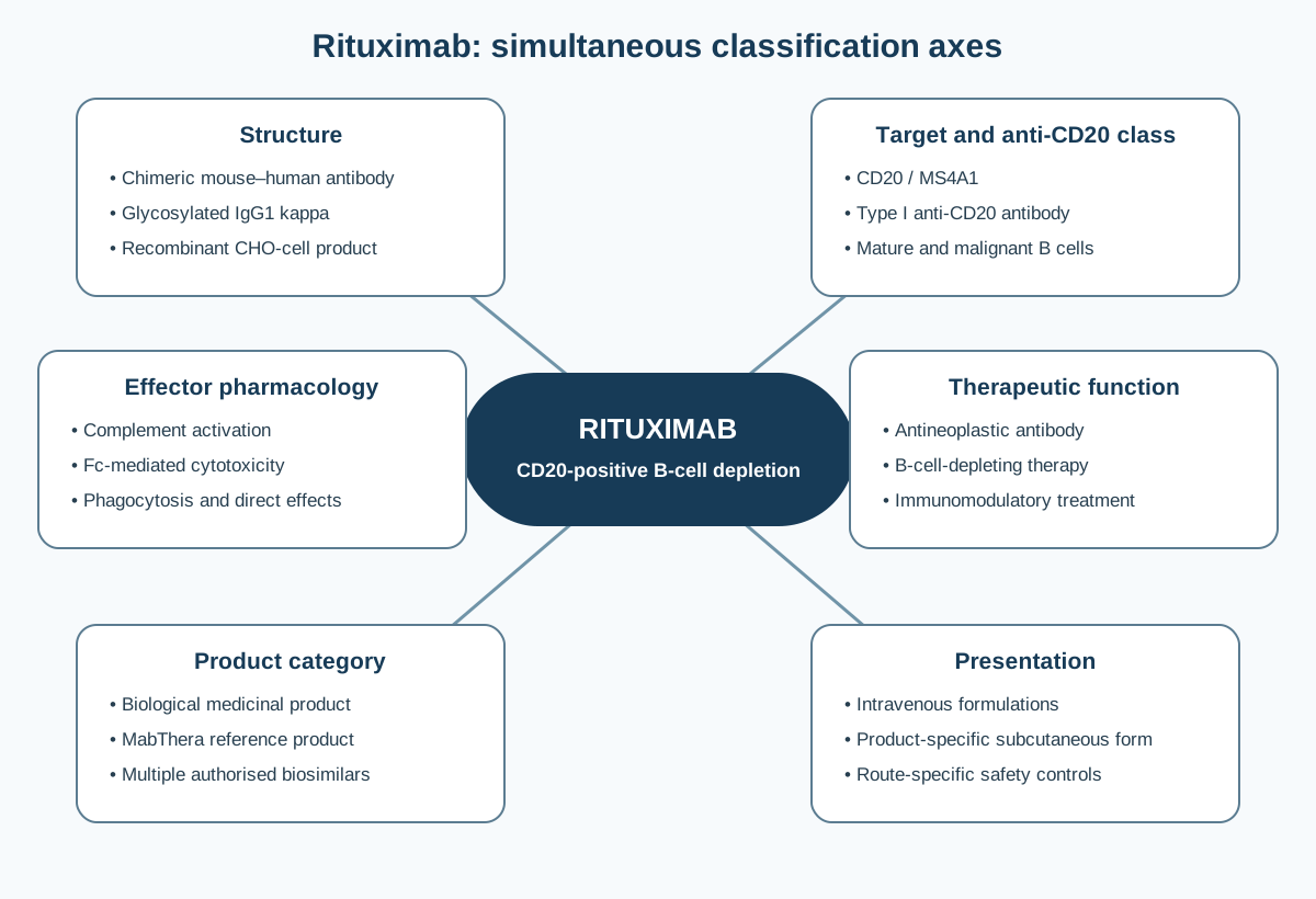
Figure 1. Rituximab occupies several simultaneous classifications. These axes are complementary rather than mutually exclusive: molecular structure enables effector function, target biology defines the affected cell population, and regulatory product/formulation status determines product-specific use and traceability.
Structural classification
Rituximab is a chimeric monoclonal antibody with murine variable regions joined to human IgG1 heavy-chain and kappa light-chain constant regions. The variable domains recognise an extracellular epitope of CD20. The Fc region recruits human immune effectors and complement. It is therefore inaccurate to describe rituximab merely as “humanised”: chimeric and humanised antibodies are structurally distinct categories.
As a glycoprotein made in living cells, rituximab is not defined solely by its amino-acid sequence. Higher-order structure, glycosylation, charge variants, aggregation, biological activity and process-related impurities are controlled attributes. An authorised biosimilar is required to demonstrate high similarity to the reference medicine through a stepwise comparability exercise; it is not assessed as a simple generic copy.
Functional anti-CD20 classification
Anti-CD20 antibodies are often divided into type I and type II agents according to experimentally observed patterns of CD20 organisation and effector activity. Rituximab is the archetypal type I antibody. Type I binding promotes redistribution of CD20 into detergent-resistant membrane microdomains and is associated with efficient complement activation. This classification is useful but not absolute: experimental assay conditions, target-cell properties and antibody concentration influence observed effects.
Biological distribution of the target
CD20 is expressed across much of B-cell development, from late pre-B cells through mature and memory B cells, and on many B-cell malignancies. It is generally absent from haematopoietic stem cells and pro-B cells, allowing regeneration of the B-cell compartment after depletion. Terminally differentiated plasma cells usually do not express CD20, so existing plasma-cell populations and pre-existing antibody production are not immediately eliminated. Nevertheless, repeated depletion can impair replenishment of antibody-producing cells and may contribute to clinically important hypogammaglobulinaemia.
Why CD20 became a therapeutic target
CD20 has several characteristics that made it attractive for antibody therapy: relatively lineage-restricted expression, high expression on many malignant B cells, proximity of its extracellular loops to the membrane, and limited shedding or release as a soluble antigen. Its persistence at the cell surface permits an antibody-coated cell to remain accessible to complement and Fc-receptor-bearing effectors. Target selection also offered a tolerable biological trade-off: mature B cells could be depleted while stem cells and early precursors retained the potential to rebuild the compartment.
This target logic does not mean that CD20 is biologically dispensable or that depletion is harmless. B cells act as antibody precursors, antigen-presenting cells, cytokine producers and organisers of immune responses. Clinical benefit in autoimmune disease can therefore reflect interruption of several B-cell functions, while adverse consequences can include impaired new antibody responses, altered immune memory and infection susceptibility.
Discovery and development history
From hybridoma technology to a therapeutic lineage marker
The development of rituximab followed the emergence of monoclonal-antibody technology and the identification of differentiation antigens on lymphocytes. Early murine antibodies could recognise tumour-associated targets, but repeated therapeutic use was constrained by immunogenicity, limited interaction with human effector systems and manufacturing challenges. Chimeric design offered a pragmatic solution: retain murine antigen-binding regions while replacing most of the molecule with human constant regions.
CD20 was pursued because it was expressed on most mature B cells and many B-cell non-Hodgkin lymphomas, but not on stem cells or most plasma cells. IDEC Pharmaceuticals developed the chimeric antibody IDEC-C2B8, later named rituximab. The programme combined target selection, an IgG1 Fc capable of recruiting immune effectors, and clinical evaluation in relapsed indolent lymphoma.
Establishing clinical efficacy
Early phase studies showed rapid peripheral B-cell depletion and antitumour activity. A pivotal multicentre study in 166 patients with relapsed low-grade or follicular lymphoma used four weekly intravenous infusions of 375 mg/m². The reported overall response rate was 48%, including complete and partial responses, and the safety profile was dominated by infusion-associated reactions, particularly during the first administration. These results were striking for a single biological agent in previously treated lymphoma.
The US Food and Drug Administration approved rituximab on 26 November 1997 for relapsed or refractory CD20-positive low-grade or follicular B-cell non-Hodgkin lymphoma. It was the first monoclonal antibody approved for cancer treatment. The European Union granted MabThera a marketing authorisation on 2 June 1998.
From single-agent lymphoma therapy to combination treatment
Rituximab's subsequent development established combination regimens with chemotherapy and broadened its role across follicular lymphoma, diffuse large B-cell lymphoma and chronic lymphocytic leukaemia. These advances also complicated causal attribution. Cytopenias, infection, tumour lysis, cardiac events and fatal outcomes may arise from rituximab, the malignancy, tumour burden, chemotherapy, corticosteroids, prior transplantation or their interaction. Pharmacovigilance therefore requires regimen-level and patient-level context rather than isolated event counting.
Expansion into autoimmune disease
The therapeutic hypothesis in rheumatoid arthritis was that B cells contribute to autoantibody production, antigen presentation and inflammatory signalling even when long-lived plasma cells are not directly targeted. Clinical development led to authorised use in severe active rheumatoid arthritis in defined patients, in combination with methotrexate.
Rituximab later gained EU indications in granulomatosis with polyangiitis and microscopic polyangiitis, where B-cell depletion affects pathogenic immune pathways associated with antineutrophil cytoplasmic antibodies, and in moderate to severe pemphigus vulgaris, an autoantibody-mediated blistering disorder. These indications made clear that anti-CD20 treatment is not simply “chemotherapy without chemotherapy”; it is targeted remodelling of a cellular immune compartment.
Formulation development
The original product is administered intravenously under close supervision with resuscitation capability. A subcutaneous MabThera formulation was subsequently developed for specified adult haematological indications. It contains recombinant human hyaluronidase to facilitate administration of a larger volume into subcutaneous tissue. The subcutaneous presentation is not interchangeable at the point of administration with the intravenous presentation: strengths, volumes, route, eligibility and first-dose requirements differ. Product selection and administration therefore form part of the safety system.
Biosimilar era
Loss of market exclusivity and maturation of the biosimilar pathway led to multiple authorised rituximab biosimilars in Europe. A biosimilar must demonstrate high similarity in analytical characteristics, biological activity, pharmacokinetics and clinical performance, with no clinically meaningful differences from the reference product. Authorised extrapolation of indications rests on the totality of evidence, not on repeating every reference-product efficacy trial.
Amgen's Riabni (rituximab-arrx) was approved in the United States in December 2020 as a biosimilar to Rituxan. This product context must remain jurisdiction-specific and should not be converted into an assumed EU brand identity. Where Riabni is reported, the suffix, brand, batch, indication and regimen help distinguish product attribution from the broader rituximab and anti-CD20 safety evidence.
For pharmacovigilance, biosimilar availability increases the importance of recording the exact product name and batch number. “Rituximab” alone identifies the active substance but may be insufficient for product-specific signal detection, quality-defect assessment, immunogenicity evaluation or investigation of administration errors.
CD20 biology
CD20 is a nonglycosylated, multipass membrane protein encoded by MS4A1. It has small extracellular regions, does not possess a conventional long cytoplasmic signalling domain and is thought to participate in B-cell activation and calcium regulation. Its expression and membrane organisation vary by B-cell maturation stage and disease state.
The rituximab epitope lies within the large extracellular loop. Binding is bivalent and can cluster or redistribute CD20. Because the antigen is close to the membrane and is not readily shed, deposited complement components and Fc regions are positioned near the target-cell surface. This geometry helps explain why CD20 is an effective “docking site” for immune-mediated elimination even though the complete physiological function of CD20 is not required to explain every therapeutic effect.
Target density matters. Cells with low CD20 expression may be less efficiently opsonised. Complement-regulatory proteins such as CD46, CD55 and CD59 can oppose complement injury. Fc gamma receptor expression and function, macrophage availability, natural-killer-cell function, tissue accessibility and prior therapy can all change the response. Malignant cells may also adapt through CD20 downregulation, antigenic modulation, internalisation or selection of low-expressing clones.
Detailed mechanism of action
The clinically relevant mechanism is best understood as a sequence:
- rituximab distributes to accessible compartments and binds CD20-positive cells;
- surface-bound antibody reorganises CD20 and exposes Fc domains;
- complement proteins and Fc-receptor-bearing cells recognise the opsonised target;
- lysis, cytotoxicity, phagocytosis or direct death signals remove target cells;
- depletion alters malignant-cell burden or pathogenic B-cell functions;
- repopulation occurs from CD20-negative precursors, with timing and composition influenced by dose, repeat courses, disease and concomitant immunosuppression.
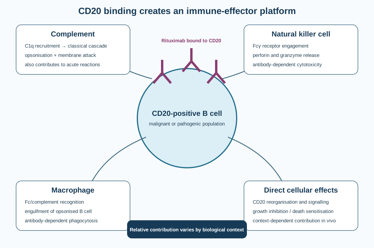
Figure 2. Rituximab coats a CD20-positive B cell and can recruit several interacting elimination pathways. The diagram does not assign a universal hierarchy: complement, natural-killer cells, macrophages and direct signalling may contribute differently across diseases and tissue compartments.
Complement-dependent cytotoxicity
When rituximab molecules bind and organise on the B-cell surface, adjacent Fc regions can recruit C1q and initiate the classical complement cascade. Complement cleavage generates opsonins and inflammatory mediators; assembly of the membrane-attack complex can disrupt the target-cell membrane. Complement activation can also contribute to acute infusion reactions through release of anaphylatoxins and cytokine amplification.
Complement-dependent cytotoxicity is strongly supported by in-vitro evidence, pharmacodynamic observations and the type I behaviour of rituximab. Its contribution in an individual patient is constrained by complement availability, target density and complement-regulatory proteins. Heavy tumour burdens may consume complement. Repeated or closely spaced exposure can therefore occur in a biological environment different from the first infusion.
Antibody-dependent cellular cytotoxicity
The Fc domain of cell-bound rituximab engages activating Fc gamma receptors on natural killer cells and other cytotoxic effectors. Natural-killer-cell activation can result in degranulation and release of perforin and granzymes, damaging the antibody-coated B cell. Fc-receptor polymorphisms and effector-cell competence have been associated with response in some settings, but such associations are not a universal clinical decision rule.
Antibody-dependent cellular phagocytosis
Macrophages and other phagocytes recognise Fc domains and complement-opsonised surfaces, engulfing rituximab-coated B cells. Experimental and translational evidence increasingly supports phagocytosis as an important mechanism of depletion, particularly in reticuloendothelial organs. This pathway also illustrates a saturable system: very high circulating target-cell burdens may tax available effector capacity.
Direct signalling and cell-death effects
Cross-linking or reorganisation of CD20 can alter membrane microdomains and intracellular signalling, inhibit proliferation, sensitise cells to cytotoxic agents or induce forms of cell death in experimental systems. Reported pathways include changes in calcium handling, kinase signalling and apoptotic machinery. Their magnitude varies markedly among cell lines and experimental conditions.
Direct cell death should therefore be described as a plausible contributing mechanism, not as the sole or uniformly dominant explanation for clinical efficacy. Statements that rituximab simply “induces apoptosis” erase the better-established role of host immune effectors and the uncertainty about relative contributions in vivo.
B-cell depletion and disease modification
Peripheral B cells often fall rapidly after rituximab exposure, but blood counts are an incomplete surrogate for tissue depletion. Lymph nodes, spleen, bone marrow and inflamed tissues differ in antibody penetration, target-cell phenotype and effector-cell access. A patient can have profound peripheral depletion while retaining tissue B-cell populations.
In B-cell malignancy, benefit follows elimination of malignant CD20-positive cells and enhancement of chemotherapy-mediated control. In autoimmune disease, depletion can reduce antigen presentation, inflammatory cytokine networks, activation of autoreactive T cells and generation of short-lived plasmablasts. Long-lived plasma cells are generally spared, explaining why pre-existing antibodies do not disappear immediately. Clinical improvement may lag behind depletion and need not correlate directly with the speed of peripheral repopulation.
Repopulation usually begins with immature or naïve B cells before memory compartments fully recover. Repeat treatment, cumulative immunosuppression and patient-specific immune reserve can produce delayed or incomplete recovery. This is the biological bridge between mechanism and the later risks of impaired vaccine response, hypogammaglobulinaemia and infection.
Clinical-use architecture
The authorised EU indications and regimens are product- and formulation-specific and evolve through regulatory procedures. The current MabThera product information should be consulted before use. At a conceptual level, its authorised uses fall into two domains.
| Domain | Major EU-authorised disease groups | Therapeutic logic | Important PV context |
|---|---|---|---|
| Haematological malignancy | Follicular lymphoma, diffuse large B-cell lymphoma, chronic lymphocytic leukaemia | Remove malignant CD20-positive B cells, commonly with chemotherapy | Tumour burden, tumour lysis, cytopenias, infection, chemotherapy interaction |
| Autoimmune/inflammatory disease | Rheumatoid arthritis, granulomatosis with polyangiitis, microscopic polyangiitis, pemphigus vulgaris | Interrupt pathogenic B-cell functions and autoantibody-generating pathways | Repeated courses, corticosteroids/immunosuppressants, chronic infection risk, PML education and patient alert card |
Regimens vary substantially across diseases. Oncology schedules may be linked to chemotherapy cycles or maintenance treatment; autoimmune schedules commonly use treatment courses separated by clinical reassessment. Dose, interval and co-medication cannot safely be inferred from the active substance name.
Pharmacokinetics and pharmacodynamics
Rituximab pharmacokinetics are nonlinear and time-dependent in some populations because CD20-positive cells form a target-mediated clearance pathway. Patients with a high circulating or tissue B-cell burden may clear antibody more rapidly early in treatment. As tumour burden falls and target sites become less abundant, systemic exposure and half-life may increase. Body size, sex, disease, tumour burden, treatment cycle and anti-drug antibodies can contribute to variability.
Following intravenous administration, exposure is systemic immediately. Subcutaneous administration has an absorption phase and uses a fixed, indication-specific dose after eligibility requirements have been met. Pharmacokinetic comparability supported the subcutaneous development programme, but the routes have different local-reaction and medication-error considerations.
Pharmacodynamics include rapid peripheral CD20-positive B-cell depletion and later repopulation. B-cell counts, immunoglobulin concentrations, disease biomarkers and clinical response describe different aspects of effect and should not be treated as interchangeable.
Safety profile as a consequence of mechanism and context
Rituximab safety is shaped by four interacting layers:
- acute target-cell and immune-effector activation;
- consequences of malignant-cell destruction;
- sustained or repeated B-cell depletion;
- the underlying disease and concomitant treatment.
Infusion-related reactions
Infusion-related reactions are most frequent with the first intravenous infusion. Fever, chills, rigors, rash, pruritus, dyspnoea, bronchospasm, hypotension, angioedema and other manifestations may occur. Severe reactions can include hypoxia, pulmonary events, arrhythmia, myocardial ischaemia, shock or death.
The mechanism may involve cytokine release, complement activation and rapid interaction with a large burden of circulating target cells. Risk is therefore not equivalent to conventional IgE-mediated allergy, although true hypersensitivity can occur. Premedication, controlled initial infusion rates, close observation, availability of resuscitation facilities and protocol-directed interruption or discontinuation are risk controls specified in product information.
Subcutaneous administration can cause local and systemic administration-related reactions. The requirement for a preceding full intravenous dose is an important product-specific control because it places the initial exposure in a setting where the rate can be adjusted or stopped immediately.
Tumour lysis syndrome
Rapid destruction of a large malignant-cell burden can release intracellular potassium, phosphate and nucleic-acid metabolites, producing hyperkalaemia, hyperphosphataemia, hypocalcaemia, hyperuricaemia, acute kidney injury, arrhythmia, seizure or death. Tumour lysis is a disease- and burden-dependent risk amplified by effective cytoreduction, not a generic manifestation of B-cell depletion.
Risk assessment, hydration, biochemical monitoring and urate-lowering strategies should follow the relevant product information and oncology protocols. Safety reports should capture baseline tumour burden, concomitant cytotoxic therapy, prophylaxis, temporal relationship and laboratory evolution.
Infections and viral reactivation
B-cell depletion can impair humoral responses and interact with chemotherapy, corticosteroids, prior transplantation or other immunosuppressants. Serious bacterial, viral, fungal and opportunistic infections have been reported. The individual contribution of rituximab may be difficult to isolate, but mechanistic plausibility and accumulated clinical evidence make infection surveillance central to its lifecycle safety management.
Hepatitis B virus reactivation is a particularly important preventable risk. Reactivation can occur in patients with current infection and in those with evidence of resolved infection, sometimes leading to fulminant hepatitis and death. Screening before treatment, expert assessment, prophylaxis or monitoring as appropriate, and continued vigilance after treatment are necessary because reactivation may be delayed.
Rituximab has also been associated with progressive multifocal leukoencephalopathy (PML), a rare, often fatal demyelinating disease caused by JC-virus reactivation. New neurological, cognitive or psychiatric symptoms require urgent evaluation. Diagnostic delay is a critical failure mode because symptoms may initially be attributed to malignancy, autoimmune disease, infection, stroke, treatment toxicity or ageing.
Hypogammaglobulinaemia and impaired vaccine response
Plasma cells are not directly depleted, but repeated removal of precursor and memory B-cell populations can impair replenishment of antibody-producing cells. Immunoglobulin concentrations may decline, particularly after repeated courses or in patients with low baseline levels, prior immunosuppression or haematological malignancy. Persistent hypogammaglobulinaemia can be accompanied by recurrent or severe infection.
Baseline and longitudinal immunoglobulin assessment is clinically valuable in risk-based care, especially where repeated courses are planned. The precise monitoring schedule and response to abnormalities depend on indication, product information, local guidance and patient context. Safety evaluation should distinguish an isolated laboratory decrease from clinically consequential immune deficiency.
Rituximab can blunt responses to non-live vaccines. Vaccination planning should occur before B-cell-depleting therapy when feasible, and live vaccines require particular caution according to product information and immunisation guidance. A poor serological response after treatment is an expected pharmacological consequence, not proof that a vaccine product failed.
Cytopenias
Neutropenia can occur during treatment or after a delay. Late-onset neutropenia is generally described weeks to months after the final dose and may be detected incidentally or during an infection. Its mechanism is not fully established; proposed explanations include altered marrow homeostasis during B-cell recovery, immune-mediated processes and changes in cytokine networks. Attribution is complicated by chemotherapy, marrow disease, infection and other medicines.
Anaemia, thrombocytopenia and other cytopenias also occur in relevant populations. Reports require dates, serial counts, marrow status, co-therapy, disease activity, infection and dechallenge/rechallenge information.
Severe mucocutaneous reactions
Rare severe mucocutaneous reactions, including fatal cases, have been reported. Phenotypes may include Stevens–Johnson syndrome, toxic epidermal necrolysis, paraneoplastic pemphigus and other bullous disorders. The differential diagnosis is difficult in patients with malignancy, autoimmune blistering disease or multiple concomitant medicines. Detailed dermatological morphology, mucosal involvement, biopsy findings, latency and alternative causes are essential for meaningful case assessment.
Cardiovascular, pulmonary, renal and gastrointestinal events
Serious cardiovascular reactions can occur during administration, particularly in patients with pre-existing cardiac disease or cardiotoxic co-therapy. Pulmonary events may be acute components of an infusion reaction or delayed inflammatory/toxic syndromes. Renal injury may occur with tumour lysis or other clinical complications.
In rheumatoid arthritis, gastrointestinal perforation has been reported in treated populations, but underlying disease and concomitant corticosteroids or nonsteroidal anti-inflammatory drugs may contribute. Pharmacovigilance assessment must avoid converting temporal association into a single-cause conclusion.
A pharmacovigilance model for rituximab
The safety system must connect product identity, indication, regimen, immune status and time. A report of “infection after rituximab” is only the beginning of an assessment.
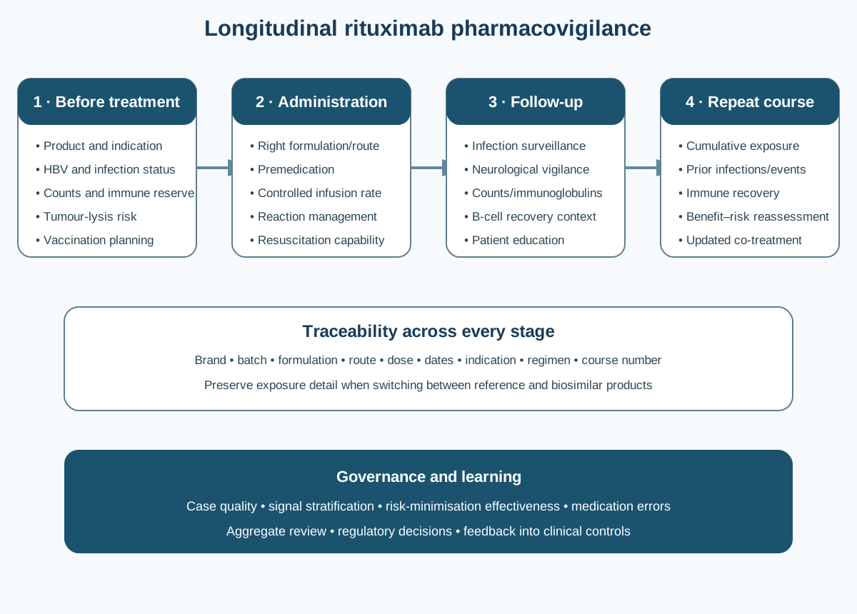
Figure 3. Rituximab pharmacovigilance spans pre-treatment risk characterisation, administration controls, longitudinal immune surveillance and product-level traceability. Controls vary by indication and product information; the diagram is a governance framework, not a dosing protocol.
Minimum case context
High-value case documentation includes:
| Domain | Information needed |
|---|---|
| Product | Brand/trade name, active substance, formulation, route, strength, batch number, country and source |
| Exposure | Dose, dates, infusion number, course number, infusion rate, interruptions, premedication and prior rituximab exposure |
| Indication | Exact disease, stage/activity, tumour burden and relevant organ involvement |
| Co-treatment | Chemotherapy regimen, corticosteroids, immunosuppressants, growth factors, antimicrobial prophylaxis and recent vaccines |
| Baseline immune status | Hepatitis B markers, relevant infection screening, immunoglobulins, blood counts, prior infections and prior immune-depleting therapy |
| Event | Onset, phenotype, severity, investigations, treatment, outcome, dechallenge and rechallenge |
| Competing causes | Underlying disease, transplantation, marrow involvement, procedures, exposure to infection and other suspect medicines |
Time-to-onset is mechanism-dependent
Acute infusion reactions usually cluster during or shortly after administration. Tumour lysis follows rapid malignant-cell destruction. Cytopenias may be early or delayed. Viral reactivation, PML, hypogammaglobulinaemia and serious infection may emerge months after a treatment course. A surveillance window limited to the administration day will therefore systematically miss important risks.
Signal detection challenges
Confounding by indication and regimen
Cancer and autoimmune disease carry different background risks. Oncology patients may have marrow suppression, tumour-related immune dysfunction and multi-agent chemotherapy; vasculitis patients may receive high-dose glucocorticoids or other immunosuppressants. Crude comparison of reporting rates across indications or products can therefore be misleading.
Channeling and switching
Patients at higher risk or with previous treatment failure may be channelled to particular regimens. Switching among reference and biosimilar products can obscure exposure if only the INN is recorded. Analyses should account for brand, batch, route, indication, line of therapy and prior exposure where data permit.
Long latency
Delayed neutropenia, hypogammaglobulinaemia, infection, hepatitis B reactivation and PML may occur after routine follow-up has shifted away from the infusion service. Linkage across oncology, rheumatology, dermatology, nephrology, neurology, infectious disease, pharmacy and primary care is therefore important.
Clinical heterogeneity
“Infusion reaction,” “infection” and “cytopenia” are broad categories. Signal evaluation improves when cases are phenotyped: timing within the infusion, affected organs, microbiological diagnosis, immune measures, grade, treatment and outcome. Grouping biologically distinct events can conceal a pattern.
Risk minimisation and evidence of effectiveness
Risk minimisation is not complete when a warning is placed in product information. A mature system asks whether the control reliably changes practice.
Before treatment
Controls include verifying the indication and product, screening for hepatitis B as specified, reviewing active infection, obtaining relevant blood counts and immune measures, assessing tumour-lysis risk, reviewing vaccination status and documenting prior anti-CD20 exposure. For repeat courses, cumulative immune effects matter more than a snapshot taken before the first-ever dose.
During administration
The correct formulation, route and dose must be independently verified. Premedication and infusion-rate instructions should be followed. Staff must recognise and grade reactions, know when to interrupt or stop treatment and have immediate access to resuscitation facilities. For subcutaneous MabThera, eligibility and prior intravenous exposure must be verified.
After treatment
Follow-up should reflect the long pharmacodynamic tail. Patients and clinicians need clear instructions about infection, neurological symptoms and delayed blood abnormalities. Repeat-course decisions should integrate clinical response, B-cell recovery where relevant, immunoglobulin trends, infections and concomitant immunosuppression.
Additional risk-minimisation measures in the EU
EMA public information describes educational material for physicians and patients when MabThera is used in rheumatoid arthritis, granulomatosis with polyangiitis, microscopic polyangiitis or pemphigus vulgaris, focused on infection risk including PML. Patients in these settings receive an alert card to carry. Physicians administering the subcutaneous product also receive material intended to minimise improper use and medication errors.
Effectiveness evidence can include documented distribution, receipt and understanding of materials; hepatitis B screening completion; alert-card provision; correct route and formulation selection; adherence to first-dose requirements; and timeliness of investigation for neurological symptoms. These are process measures and should be complemented by outcome and case-quality review.
Important pharmacovigilance topics
Product traceability and biosimilars
EU pharmacovigilance requires biological medicines to be identifiable by product name and batch number where available. This supports attribution of suspected adverse reactions, immunogenicity clusters, quality defects and distribution-specific problems. Systems that automatically collapse all brands to “rituximab” sacrifice information that may later be essential.
Traceability does not imply that every event is caused by a product difference. A brand imbalance may instead reflect market share, indication mix, reporting behaviour, hospital contracts or incomplete recording. Product-level signals require exposure-aware analysis and, where relevant, quality investigation.
Immunogenicity
Anti-rituximab antibodies can occur and may be more frequently detected in some autoimmune populations than in lymphoma. Their clinical meaning varies: they may be transient, have no discernible effect, alter exposure, accompany infusion/hypersensitivity reactions or reduce pharmacodynamic response. Assay sensitivity, drug interference, sampling time and neutralising-antibody methods must be considered before comparing incidence among studies or products.
An immunogenicity assessment should connect laboratory findings with pharmacokinetics, B-cell depletion, clinical response and adverse reactions. A positive test alone is not a complete safety signal.
Pregnancy and lactation
Rituximab is an IgG antibody and can cross the placenta, particularly later in pregnancy. Transient B-cell depletion and lymphocytopenia have been reported in some infants exposed in utero. Pregnancy cases should document timing by gestational age, indication, dose, co-medication, maternal disease, pregnancy outcome, neonatal blood counts, infection, vaccination and B-cell recovery where available.
Rituximab has been detected at low concentrations in human milk. Clinical decisions during breastfeeding must follow current product information and consider maternal need, infant age, timing and uncertainty. PV follow-up should avoid assuming either zero exposure or inevitable harm.
Medication errors
Rituximab presents several opportunities for error: confusion between intravenous and subcutaneous formulations, incorrect strength or volume, omission of the required initial intravenous administration, incorrect infusion rate, and use of a regimen belonging to another indication. Brand diversity may add selection complexity. Error reports should capture whether the product reached the patient, clinical consequences, contributing system factors and corrective actions.
Regulatory and safety evolution
Rituximab’s safety profile was not fixed at first authorisation. Wider exposure across diseases and combinations revealed rare or delayed risks that pivotal lymphoma trials could not fully characterise.
- Infusion reactions and tumour lysis were evident early and became embedded in supervised-administration and monitoring requirements.
- Rare severe mucocutaneous reactions were recognised through post-marketing experience.
- PML reports across haematological and autoimmune populations led to strengthened warnings, professional awareness and patient education.
- Hepatitis B reactivation, including severe and fatal outcomes, prompted stronger screening and management language internationally.
- Accumulating experience with repeat courses increased attention to persistent hypogammaglobulinaemia, delayed neutropenia, impaired vaccine response and infection.
- Subcutaneous formulation development created additional route-specific medication-error controls.
- Biosimilar entry elevated brand and batch traceability as an operational PV priority.
This evolution illustrates why a biological product’s pharmacovigilance history is more than a list of label changes. It is a progressive mapping of mechanism, susceptible populations, latency, preventability and the real-world performance of risk controls.
Causality assessment by event type
Different events require different evidentiary questions.
Acute administration reaction
Assess exact timing relative to infusion or injection, rate changes, signs and symptoms, intervention, response to interruption, previous exposure, premedication and alternative causes. Cytokine-release or complement-mediated reactions may occur on first exposure and do not require prior sensitisation.
Serious infection
Document organism and site, immune status, neutrophil and lymphocyte counts, immunoglobulins, concomitant immunosuppression, prophylaxis, vaccination, time since the last and cumulative courses, and underlying disease. Consider whether rituximab contributed through B-cell depletion even when another drug produced more immediate immunosuppression.
Hepatitis B reactivation
Capture baseline HBsAg, anti-HBc and anti-HBs results; HBV DNA where tested; antiviral prophylaxis and adherence; serial liver tests and viral load; timing of immunosuppression; clinical hepatitis; treatment and outcome. Missing baseline serology can prevent classification and should trigger focused follow-up.
PML
Obtain symptom chronology, neurological examination, magnetic-resonance imaging, cerebrospinal-fluid JC-virus testing, biopsy if performed, immune status, prior and concomitant immunosuppressants, malignancy history and diagnostic alternatives. Negative early testing does not necessarily resolve a clinically evolving case; assessment belongs with appropriate specialists.
Delayed cytopenia or immune deficiency
Serial data are essential. A single low count cannot show onset, nadir, recovery or recurrence. For hypogammaglobulinaemia, capture baseline and longitudinal IgG, IgA and IgM where available, B-cell counts, infection history and any immunoglobulin replacement.
Inspection and governance perspective
An inspectable rituximab safety system should demonstrate:
- product-specific reference safety information is current and controlled;
- cases retain brand, formulation, route and batch information;
- follow-up forms are tailored to infusion reactions, PML, hepatitis B reactivation, serious infection, cytopenias and pregnancy;
- cumulative analyses stratify by indication and regimen where meaningful;
- delayed events remain within surveillance and follow-up processes;
- medical review distinguishes direct toxicity, target-mediated pharmacology, regimen interaction and disease-related risk;
- medication errors involving intravenous and subcutaneous products are specifically monitored;
- additional risk-minimisation measures have distribution and effectiveness evidence;
- biosimilar or switching analyses account for exposure and reporting artefacts;
- signals, aggregate reports, risk-management plans and product-information changes are traceable to decisions.
Common failures include coding a complex infusion syndrome without preserving chronology; accepting “rituximab” without brand follow-up; treating all infections as one signal category; omitting hepatitis B baseline status; closing PML follow-up after an initially negative test despite progressive symptoms; and evaluating a repeat-course patient without cumulative immunosuppressive history.
Practical checklist
Product and exposure
- Confirm brand, formulation, route, strength and batch.
- Record every relevant dose, course and infusion rate.
- Identify switches between reference and biosimilar products.
- Capture premedication and administration interruptions.
Patient and regimen
- Record the exact indication, disease activity and tumour burden.
- Capture chemotherapy, corticosteroids and other immunosuppressants.
- Document prior transplantation and prior B-cell-depleting treatment.
- Establish baseline blood counts, infection history and relevant immune measures.
Risk-specific controls
- Verify hepatitis B screening and the resulting management plan.
- Assess tumour-lysis risk in relevant malignancy.
- Confirm vaccination review before treatment where feasible.
- Ensure route/formulation controls for subcutaneous use.
- Provide and document required educational materials and patient alert card.
Longitudinal surveillance
- Maintain awareness of delayed infection, PML, cytopenia and immune deficiency.
- Trend immunoglobulins and blood counts according to clinical context and applicable guidance.
- Investigate recurrent infections rather than treating them as unrelated episodes.
- Carry cumulative exposure and prior safety events into repeat-course decisions.
Key takeaways
Rituximab is a chimeric, glycosylated IgG1 kappa monoclonal antibody and the archetypal type I anti-CD20 agent. Its classification spans structure, engineering, target, effector function, therapeutic purpose, formulation and regulatory product status.
Its clinical effect does not arise from one universally dominant pathway. Complement-dependent cytotoxicity, Fc-receptor-mediated cytotoxicity, phagocytosis and direct signalling interact in a context determined by target density, tissue access, tumour burden, host effectors and co-treatment.
The product’s history moved from relapsed B-cell lymphoma to combination oncology regimens and then to selected autoimmune diseases. Each transition changed the treated population, exposure pattern, co-therapy and safety-surveillance problem.
Major risks include acute administration reactions, tumour lysis, serious infection, hepatitis B reactivation, PML, cytopenias, hypogammaglobulinaemia, impaired vaccine responses and rare severe mucocutaneous reactions. Their latencies range from minutes to months, requiring surveillance well beyond the infusion episode.
Biosimilars do not alter the core active-substance pharmacology, but they make reliable brand and batch capture indispensable. Formulation differences also require explicit safeguards against intravenous/subcutaneous medication errors.
The mature PV question is not simply whether an event followed rituximab. It is how product, regimen, target burden, immune reserve, cumulative exposure and risk controls interacted—and whether the resulting evidence supports a preventable pattern or a change in the benefit–risk balance.
References
- European Medicines Agency. MabThera: EPAR. Current product information, assessment history and risk-minimisation overview.
- European Medicines Agency. MabThera: EPAR product information. Current revision accessed September 2026.
- US Food and Drug Administration. Rituxan prescribing information. 2021.
- McLaughlin P, Grillo-López AJ, Link BK, et al. Rituximab chimeric anti-CD20 monoclonal antibody therapy for relapsed indolent lymphoma: half of patients respond to a four-dose treatment program. J Clin Oncol. 1998;16(8):2825–2833. doi:10.1200/JCO.1998.16.8.2825.
- Grillo-López AJ, White CA, Varns C, et al. Overview of the clinical development of rituximab: first monoclonal antibody approved for the treatment of lymphoma. Semin Oncol. 1999;26(5 Suppl 14):66–73. PMID:10561020.
- Pescovitz MD. Rituximab, an anti-CD20 monoclonal antibody: history and mechanism of action. Am J Transplant. 2006;6(5 Pt 1):859–866. doi:10.1111/j.1600-6143.2006.01288.x.
- Maloney DG, Grillo-López AJ, White CA, et al. IDEC-C2B8 (rituximab) anti-CD20 monoclonal antibody therapy in patients with relapsed low-grade non-Hodgkin's lymphoma. Blood. 1997;90(6):2188–2195.
- Smith MR. Rituximab (monoclonal anti-CD20 antibody): mechanisms of action and resistance. Oncogene. 2003;22:7359–7368. doi:10.1038/sj.onc.1206939.
- Weiner GJ. Rituximab: mechanism of action. Semin Hematol. 2010;47(2):115–123. doi:10.1053/j.seminhematol.2010.01.011.
- Beers SA, Chan CHT, French RR, Cragg MS, Glennie MJ. CD20 as a target for therapeutic type I and II monoclonal antibodies. Semin Hematol. 2010;47(2):107–114. doi:10.1053/j.seminhematol.2010.01.001.
- Klein C, Lammens A, Schäfer W, et al. Epitope interactions of monoclonal antibodies targeting CD20 and their relationship to functional properties. MAbs. 2013;5(1):22–33. doi:10.4161/mabs.22771.
- Pavlasova G, Mraz M. The regulation and function of CD20: an “enigma” of B-cell biology and targeted therapy. Haematologica. 2020;105(6):1494–1506. doi:10.3324/haematol.2019.243543.
- Athni TS, Barmettler S. Hypogammaglobulinemia, late-onset neutropenia, and infections following rituximab. Ann Allergy Asthma Immunol. 2023;130(6):699–712. doi:10.1016/j.anai.2023.01.018.
- Aksoy S, Dizdar O, Hayran M, Harputluoglu H. Infectious complications of rituximab in patients with lymphoma during maintenance therapy: a systematic review and meta-analysis. Leuk Lymphoma. 2009;50(3):357–365. doi:10.1080/10428190902730219.
- Evens AM, Jovanovic BD, Su YC, et al. Rituximab-associated hepatitis B virus reactivation in lymphoproliferative diseases: meta-analysis and examination of FDA safety reports. Ann Oncol. 2011;22(5):1170–1180. doi:10.1093/annonc/mdq583.
- Carson KR, Evens AM, Richey EA, et al. Progressive multifocal leukoencephalopathy after rituximab therapy in HIV-negative patients: a report of 57 cases. Blood. 2009;113(20):4834–4840. doi:10.1182/blood-2008-10-186999.
- Wolach O, Bairey O, Lahav M. Late-onset neutropenia after rituximab treatment: case series and comprehensive review. Medicine (Baltimore). 2010;89(5):308–318. doi:10.1097/MD.0b013e3181f2caef.
- European Medicines Agency. Truxima: EPAR. Biosimilar product overview and assessment information.
- European Medicines Agency. Riximyo: EPAR. Biosimilar product overview and assessment information.
-
European Medicines Agency. Guideline on similar biological medicinal products. CHMP/437/04 Rev 1.
-
U.S. Food and Drug Administration. Riabni (rituximab-arrx), BLA 761140: approval and current product record. Approved December 2020. https://www.accessdata.fda.gov/. Accessed 4 September 2026.
- U.S. Food and Drug Administration. Purple Book: Riabni (rituximab-arrx), BLA 761140. https://purplebooksearch.fda.gov/. Accessed 4 September 2026.
Regulatory Note
This article is an educational scientific and pharmacovigilance review, not prescribing advice. Authorised indications, dosing, formulations, contraindications, warnings, risk-minimisation measures and regulatory status vary by product and jurisdiction and may change. Consult the current product-specific Summary of Product Characteristics, package leaflet, EPAR and applicable national requirements. Statements about monitoring or operational controls are identified as product-information requirements, regulatory measures or recommended practice according to their evidentiary basis; they should not be converted into universal legal requirements without verification.