Romosozumab: Sclerostin–Wnt Biology, Bone Formation, Fracture Prevention, Cardiovascular Safety and Product Pharmacovigilance
- Romosozumab: Sclerostin–Wnt Biology, Bone Formation, Fracture Prevention, Cardiovascular Safety and Product Pharmacovigilance
- Bone remodelling from first principles
- Wnt signalling: an instruction to build
- Sclerostin: the physiological brake
- Antibody structure and pharmacological behaviour
- Why the effect is dual—and time limited
- Disease–product interplay: severe postmenopausal osteoporosis
- Therapeutic-position map: intervention point and sequence
- Pivotal development: from bone-density signal to fracture strategy
- Approval history and present standard of care
- Safety understood through biology and clinical evidence
- Evolution of the safety profile
- Official-source safety-concern framework
- Product Pharmacovigilance
- Official regulatory reference material
- Key takeaways
- References
- Regulatory Note
Romosozumab is sometimes called an anabolic osteoporosis medicine because it builds bone. That shorthand misses its most distinctive feature. It both increases bone formation and decreases bone resorption, especially early in treatment. To understand this dual action, one must first understand that bone is living tissue under continuous cellular management—not an inert mineral scaffold.
Bone remodelling from first principles
Adult bone is renewed through coordinated work by three cell families. Osteoclasts remove old or damaged bone. Osteoblasts lay down new collagen-rich matrix and mineralise it. Some osteoblasts become osteocytes, long-lived cells embedded within the mineralised matrix. Osteocytes extend fine processes through microscopic channels and sense strain, local damage and hormonal signals. They function like a distributed engineering network inside the structure they supervise.
Two related processes matter. In remodelling, osteoclast removal and osteoblast replacement occur sequentially at the same site. In modelling, formation and resorption can occur on different surfaces, allowing bone to change mass or shape without first excavating the exact site where new bone will be placed. After menopause, declining oestrogen shifts remodelling toward resorption: more bone is removed than restored. Trabeculae—the internal struts of spongy bone—may thin or disconnect, while cortical bone becomes more porous. Fracture risk then reflects not only how much mineral is measured, but also microarchitecture, prior fracture, age, falls and the speed of ongoing loss.
Wnt signalling: an instruction to build
Wnt proteins are extracellular signalling molecules. In the canonical bone pathway, a Wnt ligand binds two cooperating cell-surface proteins: a Frizzled receptor and an LRP5 or LRP6 co-receptor. A receptor converts an outside binding event into an intracellular response; a co-receptor is a required partner in the receiving complex.
Without the Wnt signal, a cellular protein complex marks beta-catenin for destruction. With Wnt bound to Frizzled–LRP5/6, that destruction machinery is restrained. Beta-catenin accumulates, enters the nucleus and helps switch on genes that favour osteoblast differentiation, activity and survival. This is not a simple on/off switch for one cell: Wnt signalling also alters communication between osteoblast-lineage cells and osteoclasts, including the balance between RANKL, which supports osteoclast formation, and osteoprotegerin, a decoy receptor that restrains it.
Sclerostin: the physiological brake
Sclerostin is a secreted glycoprotein encoded by the SOST gene and produced mainly by mature osteocytes. It binds the extracellular region of LRP5/6, with LRP4 helping its presentation, and inhibits canonical Wnt signalling. The braking analogy is useful only if kept precise: sclerostin does not destroy osteoblasts or occupy every Wnt receptor permanently. It limits the strength and availability of a signalling route that supports bone formation.
Human genetics provided a natural experiment. Loss of functional sclerostin in rare disorders such as sclerosteosis and van Buchem disease is associated with excessive bone formation and very high bone mass. These observations did not by themselves prove that temporary antibody treatment would safely prevent osteoporotic fractures, but they identified an extracellular regulator that could be reached by a biological medicine.
Antibody structure and pharmacological behaviour
Romosozumab is a humanised IgG2 monoclonal antibody. An IgG molecule contains two identical heavy chains and two identical light chains. Their paired variable regions form two matching antigen-binding sites; the constant Fc region contributes structural stability and influences persistence in circulation. “Humanised” means that most antibody sequence is human while the target-recognising regions derive from the antibody-engineering process. IgG2 is an immunoglobulin subclass generally chosen when target neutralisation is wanted without designing the medicine primarily to recruit strong immune-cell killing.
Romosozumab binds soluble sclerostin. It is therefore a neutralising antagonist of an antagonist: it does not directly stimulate LRP5/6 as a receptor agonist would. Instead, it prevents sclerostin from applying its normal extracellular restraint, allowing endogenous Wnt ligands to signal more effectively where the pathway machinery and biological context permit.
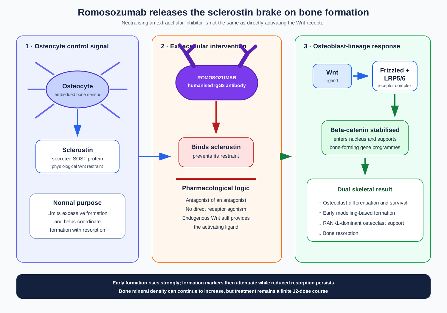
Figure 1. Osteocyte-derived sclerostin restrains the Frizzled–LRP5/6 receptor complex. Romosozumab neutralises that extracellular brake, allowing endogenous Wnt signalling to stabilise beta-catenin, support osteoblast-lineage activity and indirectly reduce osteoclast-supporting signals. The antibody is not a direct Wnt-receptor agonist.
Why the effect is dual—and time limited
Early after sclerostin inhibition, previously quiet bone surfaces can recruit lining cells and osteoblast precursors into modelling-based formation. The formation marker P1NP rises rapidly, while the resorption marker CTX falls. Biopsy data from a small FRAME substudy support predominantly modelling-based formation on cancellous and endocortical surfaces at month 2. Bone formation markers then move back toward baseline during continued dosing, whereas suppressed resorption persists through much of the 12-month course.
The attenuation is biologically important. Wnt networks contain feedback regulators, the supply and state of osteoblast-lineage cells change, and an initial burst cannot be extrapolated indefinitely. Bone mineral density can continue increasing even as the formation-marker surge wanes because less bone is being removed and earlier formation is mineralising. The approved 12-dose course reflects the studied benefit–risk programme and the waning anabolic effect; it is not evidence that osteoporosis has been cured after one year.
Disease–product interplay: severe postmenopausal osteoporosis
Osteoporosis is defined by impaired bone strength and increased susceptibility to fragility fracture. A fragility fracture follows force that would not usually break healthy bone, such as a fall from standing height. Vertebral fractures may cause pain, height loss and kyphosis, yet some are clinically silent. Hip fractures threaten mobility and independence and carry substantial downstream morbidity. A recent fracture also signals imminent risk: the probability of another fracture is especially high in the near term.
Romosozumab is therefore not positioned merely to improve a scan number. In the EU it is authorised for severe osteoporosis in postmenopausal women at high risk of fracture. The US indication is postmenopausal osteoporosis at high risk of fracture, including women with an osteoporotic fracture history or multiple risk factors, or those who failed or cannot tolerate other available therapy. Exact eligibility is jurisdiction-specific.
The product addresses a central disease imbalance by rapidly favouring formation while restraining resorption. It cannot rebuild every disconnected trabecula, reverse deformity from an established vertebral fracture, prevent falls, correct vitamin D deficiency or treat all secondary causes of bone loss. Calcium and vitamin D provide substrates for mineralisation but are not substitutes for antifracture therapy. Falls assessment, exercise, nutrition, vision, medicines that impair balance and investigation for secondary osteoporosis remain part of care.
Therapeutic-position map: intervention point and sequence
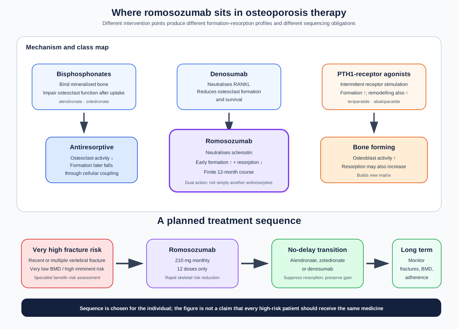
Figure 2. Osteoporosis medicines act at different points in the formation–resorption system. Romosozumab occupies a dual-action, time-limited position. For a patient at very high fracture risk, it may be used before an antiresorptive; alendronate, zoledronate or denosumab should follow without delay to preserve gains. The map describes mechanisms and sequence, not universal interchangeability.
Bisphosphonates such as alendronate and zoledronate bind bone mineral and impair osteoclast function after uptake. Denosumab neutralises RANKL and prevents osteoclast formation and survival. Both are principally antiresorptive. Teriparatide and abaloparatide intermittently stimulate the parathyroid-hormone type 1 receptor; they increase formation but also increase remodelling and resorption to varying degrees. Romosozumab releases a Wnt-related brake and has the unusual combination of increased formation and reduced resorption.
This class map explains why sequence matters. Guidelines place bone-forming therapy early for selected patients at very high fracture risk, particularly with vertebral fractures, because the immediate clinical problem is rapid risk reduction. After 12 months of romosozumab, antiresorptive treatment should begin without delay. Otherwise, bone turnover resumes and some bone-density gain can be lost. Denosumab itself also cannot be casually stopped or delayed without a planned antiresorptive transition because rebound resorption and vertebral fractures can occur; follow-on choice must therefore be planned before the first sequence begins.
Pivotal development: from bone-density signal to fracture strategy
The development programme moved from genetic and preclinical validation to dose-ranging studies, then to fracture-outcome trials. A 12-month phase 2 study in 419 postmenopausal women with low bone density compared several romosozumab regimens with placebo, alendronate and teriparatide. The 210 mg monthly regimen produced large spine and hip bone-density gains, supporting phase 3 selection. Bone density is a surrogate rather than the clinical outcome itself, so fracture trials remained essential.
FRAME: rapid benefit against placebo, then denosumab
FRAME enrolled 7,180 postmenopausal women with osteoporosis. Participants received romosozumab 210 mg monthly or placebo for 12 months; both groups then received denosumab for a further year. At month 12, new radiographic vertebral fracture occurred in 0.5% of evaluable women assigned romosozumab and 1.8% assigned placebo—a 73% relative reduction and about 1.3 percentage points absolute difference. Clinical fractures occurred in 1.6% versus 2.5%. The prespecified nonvertebral-fracture comparison was not statistically significant at 12 months.
After both groups changed to denosumab, the vertebral-fracture difference persisted through month 24. This was evidence for a treatment strategy, not proof that the first medicine remains active indefinitely: early skeletal gains were followed by a medicine that suppresses resorption. Cardiovascular events were broadly balanced in FRAME.
ARCH: active comparison and the benefit–risk tension
ARCH enrolled 4,093 postmenopausal women with osteoporosis and a fragility fracture, a population with more severe baseline skeletal risk. Participants received romosozumab monthly or alendronate weekly for 12 months; both groups then received alendronate. Through 24 months, new vertebral fracture occurred in 6.2% in the romosozumab-to-alendronate group and 11.9% in the alendronate-only group. At the later primary analysis for clinical fractures, with median follow-up of approximately 33 months, clinical fracture occurred in 9.7% versus 13.0%; nonvertebral fracture in 8.7% versus 10.6%; and hip fracture in 2.0% versus 3.2%.
These results showed that beginning with romosozumab could outperform beginning with an effective standard antiresorptive in a high-risk population. The same trial generated the central safety concern: during year 1, positively adjudicated serious cardiovascular events occurred in 2.5% with romosozumab and 1.9% with alendronate. The comparison did not establish whether romosozumab increased risk, alendronate conferred some protection, chance contributed, or several factors interacted. It did establish an imbalance too clinically important to ignore.
STRUCTURE: transition after bisphosphonates
STRUCTURE compared romosozumab with teriparatide in 436 women previously treated with oral bisphosphonates. Mean total-hip bone density increased with romosozumab and decreased initially overall with teriparatide during the 12-month comparison. The trial helps explain therapeutic positioning after prior antiresorptive exposure, but it was open-label, used bone density rather than fracture as the primary endpoint and should not be converted into a claim that one anabolic mechanism is universally superior.
Approval history and present standard of care
The cardiovascular imbalance complicated regulatory review. The US application was not approved on its first review cycle; approval followed in April 2019 with a boxed warning and a high-fracture-risk indication. In Europe, CHMP initially adopted a negative opinion in June 2019. After re-examination and a more restricted population plus risk-minimisation measures, EU authorisation was granted in December 2019.
Today romosozumab is a specialist, finite-course option rather than routine therapy for every low T-score. Its clearest place is a carefully selected postmenopausal woman whose near-term fracture danger is very high, for whom rapid skeletal benefit justifies cardiovascular uncertainty, and for whom calcium correction, monthly administration and an immediate antiresorptive sequence can be delivered reliably.
Safety understood through biology and clinical evidence
Cardiovascular events: a signal with unresolved mechanism
Myocardial infarction and stroke arise when blood supply to heart or brain is interrupted, commonly through atherosclerotic plaque disruption and thrombosis. Sclerostin and Wnt-related biology have been detected beyond bone, including in vascular tissue, but expression or genetic association is not proof that temporary antibody blockade causes an event. FDA’s multidisciplinary review concluded that there was no definitive mechanistic explanation for the clinical imbalance, while also finding no adequate alternative explanation that allowed the imbalance to be dismissed.
That distinction is central to pharmacovigilance. Mechanistic uncertainty does not cancel a randomised-trial signal, and a trial imbalance does not prove a molecular pathway. EU and US regulators translated uncertainty differently. EU product information contraindicates romosozumab in anyone with a history of myocardial infarction or stroke. The US boxed warning says not to initiate it in a patient who had myocardial infarction or stroke within the preceding year and to weigh benefit against risk in those with other cardiovascular risk factors. Both require discontinuation if myocardial infarction or stroke occurs during therapy.
Hypocalcaemia
When formation and mineralisation accelerate, calcium moves from extracellular fluid into bone. Serum calcium can therefore fall, especially if baseline calcium or vitamin D is inadequate or renal impairment limits mineral homeostasis. Pre-existing hypocalcaemia must be corrected before treatment; calcium and vitamin D should be adequate. Severe renal impairment or dialysis increases risk and warrants calcium monitoring under current product information. Paraesthesia around the mouth or limbs, cramps, spasms, confusion or seizures may indicate clinically important hypocalcaemia.
Osteonecrosis of the jaw and atypical femoral fracture
Osteonecrosis of the jaw (ONJ) is exposed jaw bone, or bone reachable through a fistula, that fails to heal in the relevant clinical setting. Antiresorptive effects can reduce remodelling needed after dental trauma, but ONJ is multifactorial: invasive dental procedures, infection, poor oral health, glucocorticoids, cancer therapy and comorbidity matter. A dental and oral-risk assessment before treatment, preventive care when appropriate and product-specific management are more useful than attributing every jaw symptom to one pathway.
An atypical femoral fracture occurs in a characteristic subtrochanteric or shaft pattern and may be preceded by dull thigh or groin pain. It has been reported rarely. Bilateral assessment may be appropriate when suspected. Neither ONJ nor atypical fracture should be inferred from anatomy alone; radiological pattern, dental history, prior and subsequent osteoporosis medicines and latency are essential.
Hypersensitivity, local reactions and immunogenicity
Rash, urticaria, angioedema and other hypersensitivity reactions can occur. Injection-site pain or erythema may instead reflect local tissue response or technique. A useful report separates local from systemic features, records time from each of the two injections, recurrence, treatment and outcome. Anti-romosozumab antibodies have been detected in a minority of treated patients; most observations do not by themselves establish altered efficacy or safety. Immunogenicity results depend on assay, timing, titre, neutralising capacity and clinical context.
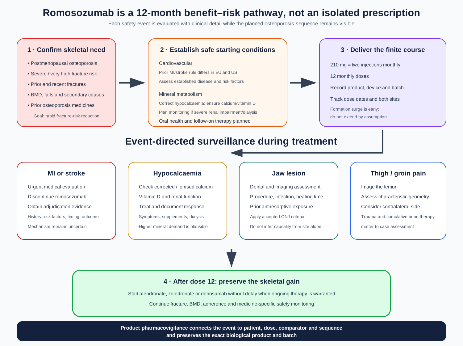
Figure 3. Safe use is a planned sequence: establish very high fracture risk, exclude or weigh key cardiovascular and metabolic risks, deliver 12 monthly doses with event-directed surveillance, then move promptly to antiresorptive maintenance. A cardiovascular event, symptomatic hypocalcaemia, jaw lesion or prodromal femoral pain triggers focused evaluation rather than automatic causal attribution.
Evolution of the safety profile
Early dose-ranging studies mainly described musculoskeletal, injection-site and laboratory effects. FRAME established antifracture efficacy and did not show the same cardiovascular imbalance later seen in ARCH. ARCH changed the regulatory trajectory by placing the imbalance against an active comparator. Regulators examined trial adjudication, baseline risk, nonclinical vascular findings and possible comparator effects; they retained cardiovascular risk as a major benefit–risk constraint rather than resolving causality.
Post-authorisation evidence has included spontaneous-report analyses, observational database studies, meta-analyses and regulator-required studies. Spontaneous databases can identify disproportionate reporting but cannot estimate incidence or remove stimulated reporting, country use patterns and confounding by indication. Observational comparisons are vulnerable to channelling because clinicians deliberately avoid romosozumab in people perceived to have greater cardiovascular risk. Meta-analyses remain limited by few events and differences among trials. Consequently, later literature has not made the authorised cardiovascular precautions obsolete.
The EU risk-management system goes beyond routine labelling. Educational material for prescribers and a patient alert card reinforce myocardial infarction and stroke, hypocalcaemia and ONJ risk management. The public RMP also includes post-authorisation studies addressing cardiovascular outcomes and other uncertainties. In the US, the boxed warning, Warnings and Precautions and Medication Guide are the principal public risk-communication tools; no romosozumab REMS is listed in current FDA REMS materials.
Official-source safety-concern framework
| Safety concern | Regulatory classification/source | Practical product-PV focus |
|---|---|---|
| Serious cardiovascular events of myocardial infarction and stroke | Important identified risk in the EU RMP; EU contraindication for any prior MI/stroke; US boxed warning and one-year non-initiation rule | Exact diagnosis and adjudication, onset, cardiovascular history and risk factors, comparator or prior therapy, action taken and outcome |
| Hypocalcaemia | Important identified risk in the EU RMP; contraindication before correction and warning in EU/US information | Baseline and event calcium, albumin, vitamin D, renal function, supplementation, symptoms and response to correction |
| Osteonecrosis of the jaw | Important potential risk in the EU RMP; warning/precaution in EU/US information | Dental procedure, infection, lesion duration, imaging, prior antiresorptives, glucocorticoids and adjudication criteria |
| Atypical femoral fracture | Labelled warning/precaution | Prodromal pain, fracture geometry, trauma, bilateral imaging, cumulative prior/subsequent bone therapy |
| Serious hypersensitivity | Labelled warning/precaution and adverse-reaction information | Timing, phenotype, systemic involvement, treatment, dechallenge and batch/product traceability |
RMP categories describe the regulator’s safety-planning framework; they are not a frequency ranking and should not be reconstructed from a package insert alone. The current public RMP, SmPC and USPI must be checked at the time of an assessment because classification and wording can change.
Product Pharmacovigilance
A case should identify romosozumab as the active substance and capture the exact product name, country, presentation, device, batch, dose, the two injection sites and administration dates. Record the clinical basis for treatment: T-scores by site, prior and recent fragility fractures, falls, secondary causes, previous osteoporosis medicines and the planned follow-on therapy. This makes benefit–risk assessment patient-specific rather than reducing the case to an adverse-event code.
For a cardiovascular report, obtain hospital records where possible: event type, diagnostic criteria, imaging, biomarkers, adjudication, onset from dose, prior MI or stroke, established cardiovascular disease, hypertension, lipids, diabetes, smoking, kidney function and concomitant medicines. Record whether treatment was stopped. Analyses should separate MI, ischaemic stroke, haemorrhagic stroke and cardiovascular death before combining them into a composite such as MACE.
For hypocalcaemia, capture corrected and ionised calcium where available, albumin, phosphate, magnesium, vitamin D, parathyroid hormone, renal function, dialysis, supplementation and symptoms. For jaw or femoral events, collect imaging, dental or surgical details, fracture morphology and cumulative exposure to bisphosphonates, denosumab and glucocorticoids. For lack of effect, determine whether the outcome was a new fracture, inadequate density response or marker change; verify adherence, dose timing, secondary osteoporosis and whether expectations exceeded what the treatment can prevent.
Aggregate surveillance should triangulate spontaneous reports, clinical and post-authorisation studies, exposure, literature and observed-to-expected analyses. Brand and batch traceability remain mandatory for a biological medicine. Most importantly, cardiovascular signal evaluation must preserve comparator, baseline risk and event adjudication: pooling unlike controls or treating reporting odds as incidence can manufacture certainty that the evidence does not contain.
Official regulatory reference material
| Official source | What it contributes |
|---|---|
| EMA romosozumab EPAR and current Product Information, revision 9 | EU indication, contraindications, warnings, adverse reactions, administration and authorisation history |
| EMA public Risk Management Plan, updated July 2026 | Current safety concerns, pharmacovigilance activities and routine/additional risk-minimisation measures |
| EMA initial Public Assessment Report | CHMP reasoning, negative opinion and re-examination, pivotal evidence, cardiovascular uncertainty and conditions of authorisation |
| Current FDA US Prescribing Information and Medication Guide | US high-risk indication, 12-dose limitation, boxed cardiovascular warning, precautions and counselling |
| FDA BLA 761062 multidisciplinary and risk-management reviews | Benefit–risk reasoning, clinical/nonclinical evaluation, safety classification and regulatory history |
| FDA REMS@FDA database | Verification of whether a product-specific REMS is currently required; none is listed for romosozumab at the date checked |
| EMA/HMA real-world study catalogue | Protocol and status information for regulator-required post-authorisation studies |
Key takeaways
Romosozumab is a humanised IgG2 antibody that neutralises sclerostin; it antagonises an extracellular inhibitor rather than directly agonising the Wnt receptor. The result is an early, largely modelling-based formation response plus reduced resorption. In selected postmenopausal women at very high fracture risk, 12 months of therapy followed by an antiresorptive can reduce fractures more than placebo-first or alendronate-first strategies studied in pivotal trials. The cardiovascular imbalance in ARCH remains a regulatory constraint despite an unresolved mechanism and mixed later evidence. Safe use is inseparable from cardiovascular selection, calcium correction, dental awareness, exact 12-dose scheduling and planned antiresorptive follow-on therapy.
References
- European Medicines Agency. Evenity: EPAR, Product Information, revision 9. Product information updated 7 May 2025; EPAR page checked 3 September 2026.
- European Medicines Agency. Evenity: Risk Management Plan. Updated 9 July 2026.
- European Medicines Agency. Evenity: EPAR Public Assessment Report. EMA/26554/2020.
- U.S. Food and Drug Administration. Evenity (romosozumab-aqqg) US Prescribing Information and Medication Guide. Current electronic label checked September 2026.
- U.S. Food and Drug Administration. BLA 761062: Multidisciplinary Review and Evaluation—romosozumab. 2019.
- U.S. Food and Drug Administration. BLA 761062: Division of Risk Management Review. 2019.
- Cosman F, et al. Romosozumab treatment in postmenopausal women with osteoporosis. N Engl J Med. 2016;375:1532–1543.
- Saag KG, et al. Romosozumab or alendronate for fracture prevention in women with osteoporosis. N Engl J Med. 2017;377:1417–1427.
- Langdahl BL, et al. Romosozumab versus teriparatide in postmenopausal women transitioning from oral bisphosphonate therapy. Lancet. 2017;390:1585–1594.
- McClung MR, et al. Romosozumab in postmenopausal women with low bone mineral density. N Engl J Med. 2014;370:412–420.
- Eriksen EF, et al. Modeling-based bone formation after 2 months of romosozumab treatment: results from FRAME. J Bone Miner Res. 2022;37:36–40.
- Delgado-Calle J, Bellido T. Role and mechanism of action of sclerostin in bone. Bone. 2017;96:29–37.
- National Osteoporosis Guideline Group. Clinical guideline for the prevention and treatment of osteoporosis. Updated December 2024.
- European Medicines Agency/HMA. Catalogue of real-world data sources and studies: romosozumab post-authorisation study records. Checked September 2026.
- U.S. Food and Drug Administration. REMS@FDA database. Checked 3 September 2026.
Regulatory Note
This educational article does not replace current authorised product information, local risk-minimisation materials or specialist clinical judgement. EU and US cardiovascular contraindications and warnings differ materially. Eligibility, calcium monitoring, dental management, administration and follow-on therapy must be verified against the current jurisdiction-specific SmPC or USPI.