Teprotumumab: IGF-1 receptor biology, thyroid eye disease, and product pharmacovigilance
- Teprotumumab: IGF-1 receptor biology, thyroid eye disease, and product pharmacovigilance
- Why this medicine is scientifically unusual
- First principles: what a receptor does
- Antibody structure and the kind of blockade involved
- Thyroid eye disease: why an eye disorder begins outside the eye
- The TSHR–IGF-1R model—and its uncertainty
- What the medicine can change—and what it cannot promise
- Indication-by-indication deep dive
- Development: from failed oncology hypothesis to tissue-directed therapy
- Where teprotumumab sits now
- Administration is part of the safety system
- Mechanism-linked safety
- Product Pharmacovigilance
- What a good benefit–risk decision looks like
- Official regulatory reference material
- References
- Regulatory Note
Why this medicine is scientifically unusual
Teprotumumab is a fully human monoclonal antibody directed against the insulin-like growth factor 1 receptor (IGF-1R). It is authorised for thyroid eye disease (TED), an autoimmune disorder in which tissues inside the bony orbit become inflamed and enlarged. The eye can be pushed forward, eye movements can become restricted, the cornea may be exposed, and—in the most dangerous cases—the optic nerve can be compressed.
The unusual part is the route by which the medicine arrived. IGF-1R was first pursued as a cancer target because it transmits growth and survival signals. Teprotumumab, then called R1507, did not become an oncology medicine. Investigators later connected IGF-1R signalling to the behaviour of orbital fibroblasts in TED, and the antibody was repurposed. The United States approved it in January 2020; the European Union authorised it in June 2025.[1–4]
That history is more than trivia. It explains both the therapeutic idea and several adverse effects: a receptor involved in orbital tissue activation is also involved in glucose physiology, fetal development, and inner-ear biology.
First principles: what a receptor does
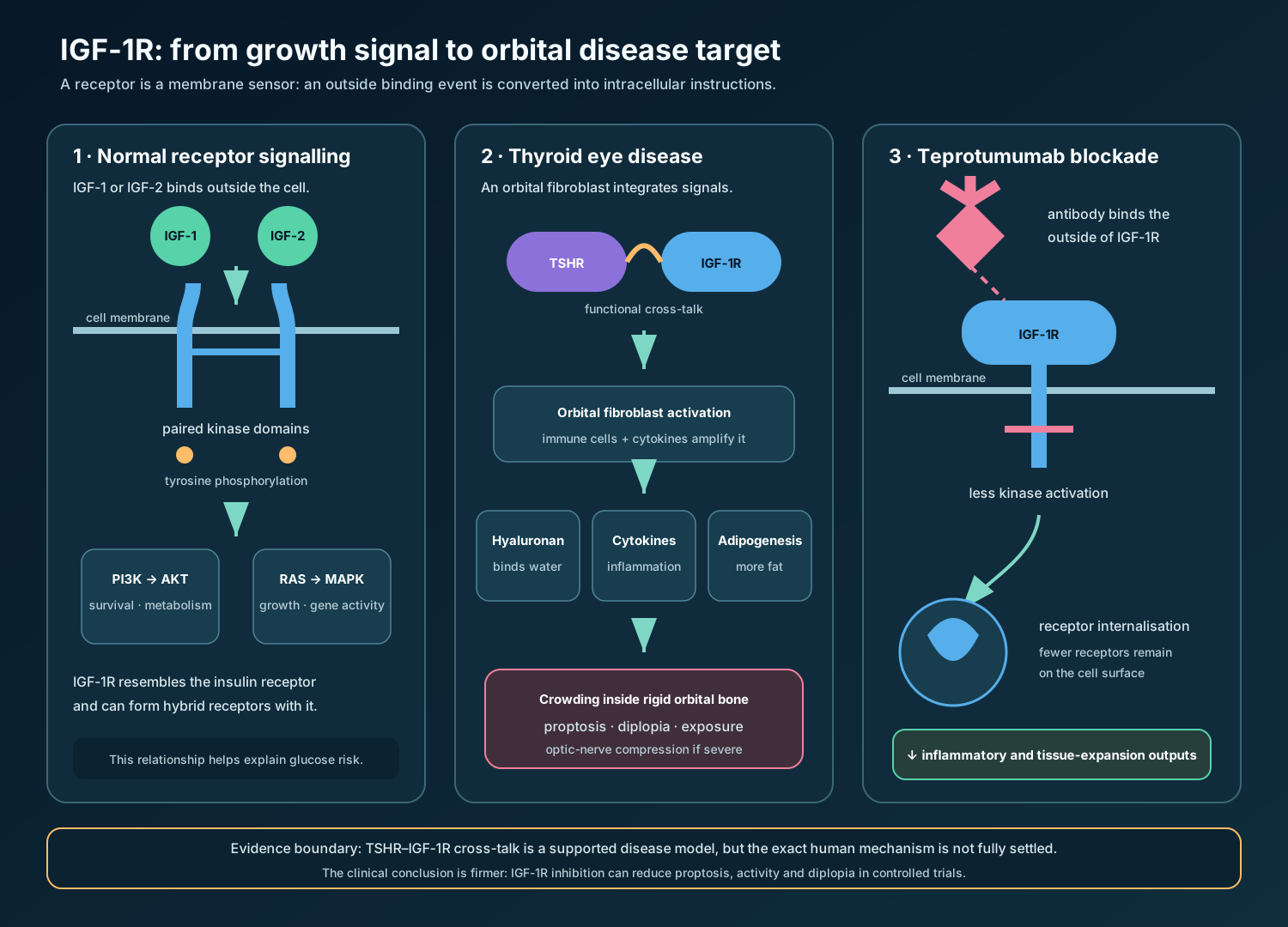
A cell membrane is not simply a wall. It is a selectively controlled boundary. A receptor is a protein that spans or sits in that boundary and converts an extracellular message into intracellular action. A useful analogy is a doorbell wired to several rooms: pressing the outside button does not enter the house, but it starts electrical signals inside. The analogy has limits—receptors can change number, pair with other receptors, and activate several molecular circuits at once—but it captures signal conversion.
IGF-1R is a receptor tyrosine kinase. “Kinase” means that its intracellular portion can transfer phosphate groups onto proteins. Phosphorylation acts like a reversible molecular annotation: it changes what a protein can bind or do. IGF-1R is assembled as two extracellular alpha subunits and two membrane-spanning beta subunits. IGF-1 or IGF-2 binds outside; the paired intracellular kinase domains then phosphorylate tyrosines.
Those phosphorylated sites recruit adaptor proteins such as insulin-receptor substrates and SHC. Two major downstream routes are:
- PI3K–AKT signalling, which supports survival, metabolism, protein synthesis, and cellular responses to nutrients;
- RAS–RAF–MEK–ERK signalling, often shortened to the MAP-kinase pathway, which influences growth, differentiation, and gene expression.
IGF-1R is closely related to the insulin receptor. Each can also form a hybrid receptor with the other. This family resemblance helps explain why blocking IGF-1R may influence glucose control even though teprotumumab does not directly target the insulin receptor.
Antibody structure and the kind of blockade involved
An antibody is a Y-shaped protein built from two identical heavy chains and two identical light chains. The tips of the Y form variable binding sites; the stem is the constant, or Fc, region. The binding sites give teprotumumab its target selectivity. They recognize an extracellular part of IGF-1R, so the antibody does not need to enter the cell.
Teprotumumab behaves principally as a receptor antagonist: it reduces ligand-driven activation rather than imitating the ligand. Binding also promotes removal of IGF-1R from the cell surface and lowers receptor abundance. Fewer functional receptors mean less opportunity to relay IGF-related signals.[5,6] This differs from an agonist, which activates a receptor, and from a depleting antibody, whose main purpose is to eliminate a target-bearing cell.
The distinction matters. Teprotumumab is not a general immunosuppressant and it does not directly remove the autoantibodies that initiate Graves-related autoimmunity. It changes how a disease-relevant tissue cell responds to signals.
Thyroid eye disease: why an eye disorder begins outside the eye
TED is most often associated with Graves disease, in which the immune system makes antibodies against the thyrotropin receptor (TSHR). TSHR normally helps regulate thyroid-hormone production. The disease is not caused simply by “too much thyroid hormone”: TED can progress when thyroid tests are abnormal, normal, or being corrected. Smoking, unstable thyroid status, and radioactive iodine in selected high-risk settings can worsen risk.[7,8]
The orbit contains the globe, extraocular muscles, fat, vessels, nerves, and connective tissue within rigid bone. There is little spare room. Orbital fibroblasts—connective-tissue cells that maintain extracellular material—can respond abnormally in TED. Immune cells and fibrocyte-like cells enter the orbit; cytokines amplify inflammation; fibroblasts produce hyaluronan, a long sugar-rich molecule that binds large amounts of water. Some fibroblasts also become fat cells. Muscles and fat consequently swell within a fixed compartment.
This gives a causal chain:
- loss of immune tolerance produces TSHR-directed autoimmunity;
- immune signals activate susceptible orbital fibroblasts;
- cytokines, hyaluronan, fat formation, oedema, and later fibrosis enlarge orbital contents;
- the eye moves forward (proptosis), muscles lose flexibility (diplopia, or double vision), eyelids retract, and surface exposure causes pain or corneal injury;
- crowding at the orbital apex can impair the optic nerve, producing dysthyroid optic neuropathy, an emergency.
Activity and severity are different axes. Activity describes current inflammation—pain, redness, swelling, and recent progression—often summarized with the Clinical Activity Score (CAS). Severity describes consequence: the degree of proptosis, diplopia, exposure, or threat to sight. A person may have severe residual proptosis after inflammation has quietened.
The TSHR–IGF-1R model—and its uncertainty
TSHR is the best-established autoantigen in Graves disease. IGF-1R appears to operate as a cooperating signalling hub in orbital fibroblasts and fibrocytes. Laboratory studies show that the two receptors can associate functionally: stimulation through TSHR can engage signalling that depends partly on IGF-1R, while IGF-1R blockade reduces AKT activation, inflammatory cytokines, and hyaluronan production.[5,6]
This is commonly called receptor cross-talk—two signalling systems influencing one another rather than behaving as isolated switches. Teprotumumab can therefore dampen outputs relevant to tissue expansion even though it does not bind TSHR.
The model should not be overstated. The importance and even the functional role of naturally occurring IGF-1R-directed antibodies remain debated, patient tissues vary, and official European information says the exact mechanism in TED is not fully characterised.[1,3] The secure clinical statement is narrower: IGF-1R inhibition changes the orbital disease phenotype in controlled trials.
What the medicine can change—and what it cannot promise
By decreasing IGF-1R signalling and receptor availability, teprotumumab can reduce proptosis, inflammatory activity, and diplopia in many treated patients. A reduction of at least 2 mm in proptosis was chosen as a clinically meaningful trial threshold because it exceeds typical measurement noise and approaches the visible effect historically sought with decompression surgery.
It does not erase the autoimmune tendency, guarantee normal eye position, reliably reverse established fibrosis, or remove the need for urgent decompression when vision is threatened. Responses can be incomplete, and disease can flare after treatment. The authorised regimen is finite, but the underlying disease is not necessarily cured.
Indication-by-indication deep dive
Active, moderate-to-severe thyroid eye disease
This is where the original controlled evidence was strongest: recent-onset, active TED with substantial inflammation and proptosis. In the phase 2 trial, the week-24 composite response—at least a 2-point CAS improvement plus at least a 2-mm proptosis reduction without corresponding worsening in the other eye—occurred in 69% of teprotumumab recipients and 20% with placebo. Separation was already visible at week 6.[9]
In the pivotal OPTIC trial, 83% of treated participants versus 10% receiving placebo achieved a proptosis response at week 24. Mean proptosis changed by −2.82 mm versus −0.54 mm. Diplopia response occurred in 68% versus 29%, and disease-specific quality of life also improved.[10] These are group results, not a prediction for an individual. The trial was relatively small, follow-up was limited, and patients with immediately sight-threatening disease were not the population in whom one should delay rescue treatment.
Why can one intervention improve several features? Proptosis, inflammatory redness, and impaired motility share a congested orbital environment. Reducing fibroblast activation and water-binding matrix can shrink tissue and relieve mechanical restriction. Established scar-like remodelling, however, is less biologically reversible.
Long-duration or low-activity disease
“Inactive” does not mean that every tissue process has stopped. A later placebo-controlled trial enrolled adults with TED lasting two to ten years and CAS no greater than 1. Mean proptosis fell 2.41 mm with teprotumumab and 0.92 mm with placebo at week 24, a between-group difference of 1.48 mm.[11] This supports a modifiable component even after obvious inflammation subsides.
It does not imply that all chronic problems respond equally. Fixed muscle fibrosis, longstanding strabismus, lid malposition, or excess skin may still require rehabilitative surgery. The European indication is adults with moderate-to-severe TED; the US indication is TED without label wording restricting activity or duration.[1,2]
Sight-threatening disease
Dysthyroid optic neuropathy and severe corneal breakdown require urgent specialist management. High-dose intravenous glucocorticoids and prompt orbital decompression when response is absent or inadequate remain the established emergency routes. Teprotumumab has plausible value and is used in selected specialist settings, but pivotal trials were not designed to establish it as a substitute for time-critical decompression.[7,8]
Development: from failed oncology hypothesis to tissue-directed therapy
IGF-1R was once considered an attractive broad cancer target. R1507 entered oncology trials but did not demonstrate sufficient benefit to continue as an anticancer programme. The molecule’s failure in one disease did not invalidate its pharmacology; it showed that receptor blockade alone was insufficient in those tumour contexts.
The repurposing logic in TED was different. Orbital fibroblasts from affected patients showed altered IGF-1R biology, and ex-vivo experiments suggested that its inhibition could interrupt TSHR-associated signalling. A small clinical programme could then test an objective anatomical endpoint—millimetres of proptosis—alongside inflammation and function.
The FDA granted breakthrough-therapy designation in 2016. Approval in 2020 relied on the phase 2 study and OPTIC, against a background in which corticosteroids treated inflammation but were much less reliable for proptosis, while radiotherapy and surgery addressed selected consequences.[4,9,10] European authorisation followed in 2025 after review of the active-disease studies and the chronic/low-activity study.[1,3]
OPTIC-X supplied useful but limited extension evidence. Among participants initially given placebo, 33 of 37 achieved a proptosis response when later treated. Small retreatment groups also included responders, but their denominators were too small to define a dependable retreatment strategy.[12] Accordingly, safety in retreated patients is treated as missing information in the European risk-management framework.[3]
Where teprotumumab sits now
There is no single universal algorithm. Availability, authorisation, cost, diabetes, bowel disease, hearing risk, fertility considerations, disease activity, and the dominant clinical problem all matter.
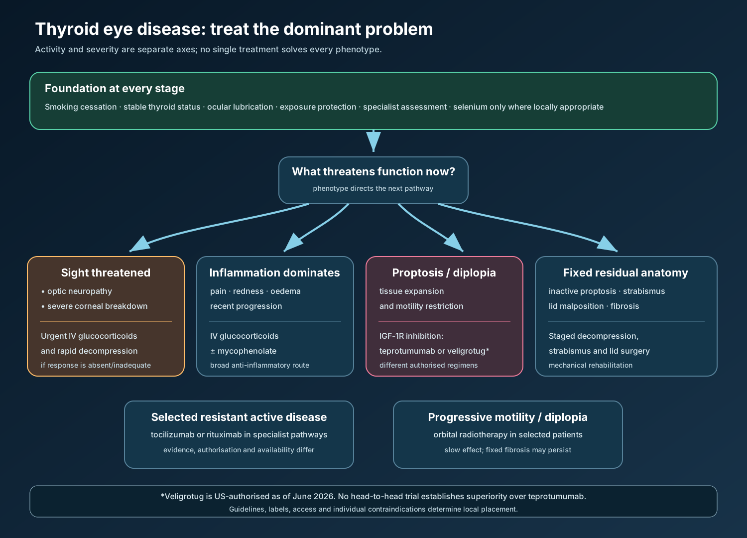
The 2021 European guideline placed intravenous methylprednisolone plus mycophenolate as first-line therapy for active moderate-to-severe disease, with teprotumumab among second-line options. The 2022 American Thyroid Association/European Thyroid Association consensus preferred teprotumumab, where available, when significant proptosis or diplopia dominates; it preferred intravenous glucocorticoids when inflammation is prominent without substantial proptosis or diplopia.[7,8] Both documents predate the 2025 EU authorisation and the 2026 US approval of veligrotug, so present-day local pathways may differ.
Therapies solve different pieces of TED:
| Approach | Principal pathway or action | Problem it addresses best | Important limit |
|---|---|---|---|
| Risk-factor control and stable thyroid status | Removes amplifiers rather than directly blocking orbital receptors | Foundational care at every stage | Does not rapidly reverse established proptosis |
| Intravenous glucocorticoids ± mycophenolate | Broad suppression of inflammatory gene expression and lymphocyte activity | Active inflammation | Limited anatomical effect on proptosis; systemic toxicity |
| Teprotumumab | IGF-1R antagonism/downregulation | Proptosis, diplopia, activity in suitable adults | Hearing, glucose, bowel and reproductive risks; incomplete/relapsing responses |
| Veligrotug | IGF-1R inhibition; five-infusion US regimen | Same therapeutic class and US indication | No head-to-head evidence proving superiority; local availability differs |
| Tocilizumab | IL-6 receptor blockade | Resistant inflammatory disease in selected patients | Not primarily an anatomical-remodelling treatment |
| Rituximab | CD20-positive B-cell depletion | Selected resistant active disease | Evidence is mixed; infusion/infectious risks |
| Orbital radiotherapy | Alters radiosensitive orbital inflammatory cells | Progressive motility restriction/diplopia | Slow effect; does not remove fixed structural disease |
| Decompression/strabismus/lid surgery | Mechanically creates space or repairs residual anatomy | Emergency optic neuropathy or inactive residual disease | Procedural risk; staged rehabilitation may be required |
Veligrotug-vvze became the second IGF-1R-targeting antibody approved for TED in the United States in June 2026. Its regimen is five infusions rather than eight.[13] Cross-trial comparisons cannot establish which antibody is more effective or safer; only a suitable comparative study could answer that.
Teprotumumab has no authorised oncology or non-TED indication. Other diseases may involve IGF-1R, but shared receptor biology is not proof of clinical benefit.
Administration is part of the safety system
The standard regimen is an intravenous loading dose of 10 mg/kg, followed by 20 mg/kg every three weeks for seven further infusions—eight infusions over approximately 21 weeks.[1,2] The first infusions are generally delivered over 90 minutes; later administration may be shortened where the applicable product information permits and earlier infusions were tolerated.
Before treatment, clinicians should document the TED phenotype, glucose status, hearing status, bowel history, pregnancy status where relevant, concomitant medicines, and the benefit expected for that patient. In Europe, audiometry is specified before treatment, around infusion 3 or 4, and after treatment. Exact product name and batch should be recorded because biological traceability links a suspected event to the administered material.[1,3]
Mechanism-linked safety
Hearing impairment
The inner ear is not passive wiring. Cochlear sensory cells, supporting tissues, neurons, and vascular structures require trophic signalling for maintenance and repair. IGF-1 signalling contributes to cochlear development and survival. Blocking IGF-1R therefore supplies a biologically plausible route to tinnitus, reduced hearing, altered sound perception, or sensorineural loss, although the precise human mechanism and susceptibility factors are not fully resolved.
The clinical phenotype is heterogeneous. Reports include tinnitus, subjective muffling, sensorineural hearing loss, autophony (one’s own voice or breathing sounds unusually loud), and symptoms compatible with a patulous Eustachian tube. Some events resolve; severe or permanent impairment has occurred. In pooled European trial data, hearing-related events occurred in 13.8% of treated participants.[1,3]
Symptoms alone are an insensitive surveillance tool because measurable threshold change can precede a complaint. Baseline and interval audiometry create an objective comparator. European information advises monitoring hearing changes for six months after treatment and considering longer follow-up when affected; clinically important loss may require discontinuation. Concurrent ototoxic medicines deserve particular scrutiny.[1]
Hyperglycaemia
IGF-1R and the insulin receptor are relatives that share intracellular pathways and can form hybrid receptors. IGF-1R antagonism may reduce insulin sensitivity or alter the balance of this network; stress and glucocorticoid exposure can add further pressure. Hyperglycaemia is therefore more likely to become clinically important in people with diabetes or impaired glucose tolerance, but it can also appear without known diabetes.
Glucose and glycated haemoglobin should be assessed before therapy, hyperglycaemia controlled before dosing, and glucose monitored during treatment. European information extends monitoring for six months after completion.[1–3] A rise in glucose is not merely a laboratory abnormality: sustained severe elevation can cause dehydration, infection risk, or acute metabolic decompensation.
Inflammatory bowel disease
IGF signalling participates in epithelial integrity and tissue repair, offering a plausible—though unproven—reason that blockade could destabilise bowel inflammation. Exacerbations of inflammatory bowel disease (IBD) are an identified risk. In 2025, US labelling expanded the warning to include IBD occurring in people without a previous diagnosis.[2] New persistent diarrhoea, abdominal pain, rectal bleeding, urgency, fever, or weight loss requires evaluation rather than automatic attribution to a mild infusion-related effect.
Embryo-fetal toxicity
IGF-1R is essential to normal growth and development. Developmental abnormalities occurred in non-human-primate studies, so pregnancy exposure is not an abstract theoretical concern. Pregnancy must be excluded as specified locally, and effective contraception is required during treatment and for six months after the last dose. European product information contraindicates use in pregnancy.[1–3]
Infusion reactions and other adverse reactions
Infusion reactions occur during or soon after intravenous administration because a large protein can trigger transient mediator release or hypersensitivity-like physiology. Manifestations may include increased blood pressure, feeling hot, rapid heartbeat, shortness of breath, headache, or muscle pain. European information reports reactions in about 4% of treated patients and requires observation throughout infusion and for 90 minutes afterwards.[1] Slowing or stopping the infusion and providing symptomatic treatment depends on severity; a severe reaction changes the risk of further exposure.
Frequently reported adverse reactions also include muscle spasm, nausea, alopecia, diarrhoea, fatigue, dry skin, altered taste, headache, weight decrease, and nail disorders.[1,2] Their mechanisms are not equally established. A mechanism-informed article should not turn every plausible biological story into a fact.
Product Pharmacovigilance
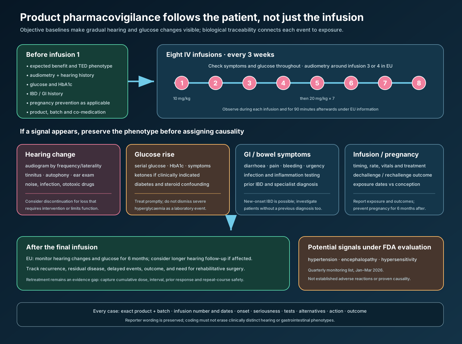
Pharmacovigilance asks four connected questions: what happened, when did it happen relative to exposure, what alternative explanations exist, and what action should follow? For teprotumumab, longitudinal measurements are especially valuable because hearing and glucose can change gradually.
Safety-concern framework
The European public assessment identifies the following risk-management categories at authorisation.[3] These categories are regulatory tools, not a ranking of frequency or seriousness.
| Category | Safety concern | Practical surveillance implication |
|---|---|---|
| Important identified risk | Hyperglycaemia | Baseline glucose/HbA1c; control pre-existing disease; monitor during and for six months after treatment in the EU |
| Important identified risk | Exacerbation of IBD | Establish bowel history; investigate new or worsening inflammatory symptoms; interrupt/discontinue according to severity and label |
| Important identified risk | Infusion-related reactions | Observe during and after every infusion; record timing, rate, treatment, and rechallenge outcome |
| Important identified risk | Hearing impairment | Baseline and serial audiometry; symptom review; assess ototoxic co-medication; follow after treatment; consider discontinuation for consequential loss |
| Important potential risk | New-onset IBD | Do not restrict vigilance to patients with known IBD; the US warning was strengthened in 2025 |
| Important potential risk | Embryo-fetal toxicity | Pregnancy prevention, testing as applicable, exposure reporting and outcome follow-up |
| Missing information | Safety in retreated patients | Treat repeat courses as an evidence gap; document cumulative exposure and outcomes carefully |
Current risk-management plans can evolve. The latest national/EU product information and current RMP summary should always supersede a historical assessment table.
A case-capture minimum dataset
For any suspected adverse reaction, collect:
- exact active substance and product name, batch/lot number, dose, infusion number, infusion dates and rate;
- indication, disease duration/activity, age, relevant history, and concomitant medicines;
- event onset, seriousness, clinical description, diagnostic tests, treatment, outcome, dose interruption, and rechallenge;
- hearing: baseline and post-event audiograms by frequency, laterality, tinnitus/autophony symptoms, ear examination, noise exposure, infection, prior loss, and ototoxic medicines;
- hyperglycaemia: baseline HbA1c/glucose, diabetes therapy, steroid exposure, serial values, ketones when indicated, symptoms, and treatment change;
- gastrointestinal events: prior IBD, stool frequency, bleeding, inflammatory markers, imaging/endoscopy, infection studies, and specialist diagnosis;
- pregnancy exposure: dates relative to conception, contraception failure, maternal course, pregnancy outcome, and infant follow-up within consent and local requirements.
Reporter terms should be retained, then coded using the current MedDRA version. “Hearing changed” must not be collapsed prematurely into a diagnosis: tinnitus, conductive loss, sensorineural loss, and patulous Eustachian-tube dysfunction are clinically different phenomena.
Expectedness, seriousness and causality
Seriousness is determined by regulatory outcome criteria—death, life threat, hospitalisation, disability, congenital anomaly, or another medically important condition—not by how dramatic a symptom sounds. A permanent hearing deficit may be serious because it causes disability even without hospitalisation.
Expectedness is judged against the applicable reference safety information for the reporting jurisdiction and period. A labelled event can still be reportable and signal-relevant when its severity, latency, permanence, dose pattern, or susceptible population differs from current knowledge.
Causality should integrate timing, biological plausibility, alternative causes, dechallenge, rechallenge, and objective data. TED itself can affect quality of life but does not usually explain a new audiometric threshold shift. Diabetes, glucocorticoids, infection, age-related loss, noise, bowel pathogens, and other medicines may confound individual cases without erasing a consistent aggregate pattern.
Signal detection and regulatory evolution
Hearing risk illustrates why approval is the beginning of safety characterisation. Hearing events were seen in development and discussed during the 2019 FDA advisory review, but the range and possible permanence became clearer with broader use. FDA adverse-event monitoring contributed to a July 2023 label update strengthening warnings and calling for hearing assessment before, during, and after treatment.[14,15]
In the EU, hearing impairment is an important identified risk supported by additional educational materials for healthcare professionals and patients. Embryo-fetal toxicity also has additional risk-minimisation material.[1,3] A required post-authorisation study compares different treatment durations and retreatment and contains a hearing substudy; it is intended to address exposure and repeat-course uncertainty.[3]
The FDA’s January–March 2026 quarterly safety communication listed hypertension, encephalopathy, and hypersensitivity among potential signals under evaluation for teprotumumab.[16] Inclusion in that list does not establish causation, frequency, or a new adverse reaction. It means FDA identified information meriting further evaluation; the correct PV response is targeted case review and follow-up while awaiting a regulatory conclusion.
REMS status and additional risk minimisation
The original FDA review concluded that a Risk Evaluation and Mitigation Strategy (REMS) was not required; risks could be managed through prescribing information and routine pharmacovigilance.[17] The current US label does not describe a product-specific REMS. “No REMS” does not mean “no risk-management obligations.”
Europe uses additional educational materials focused on hearing impairment and embryo-fetal toxicity, alongside the SmPC, patient leaflet, additional-monitoring symbol, routine adverse-reaction reporting, and post-authorisation studies.[1,3] These systems differ in form, so local implementation must follow the authorised jurisdiction rather than importing one region’s requirements into another.
What a good benefit–risk decision looks like
The best candidate has a meaningful, measurable TED problem that IGF-1R inhibition can plausibly improve and for whom hearing, metabolic, bowel, and reproductive risks can be assessed and monitored. The treatment objective should be explicit—for example, reduce proptosis enough to improve exposure and appearance, or improve diplopia and function—not simply “treat TED.” Baseline photographs, exophthalmometry, motility/diplopia assessment, CAS, quality-of-life impact, audiometry, and metabolic data make later benefit–risk reassessment concrete.
After treatment, improvement does not remove the need for follow-up. Recurrence, residual fibrosis, delayed hearing change, glycaemic deterioration, and staged rehabilitative surgery remain possible. A finite infusion course creates a defined exposure period, not a defined end to disease or surveillance.
Official regulatory reference material
The following are the primary materials for current product pharmacovigilance work. Always use the latest version applicable to the administered product and reporting jurisdiction.
- European Medicines Agency. Tepezza: EPAR — Product information (SmPC, package leaflet and labelling). Current page and revision history.
- US Food and Drug Administration. TEPEZZA (teprotumumab-trbw) US Prescribing Information, BLA 761143, current approved label in Drugs@FDA.
- European Medicines Agency. Tepezza EPAR Public Assessment Report, including benefit–risk assessment and risk-management discussion.
- US Food and Drug Administration. Multi-discipline review and approval package, BLA 761143.
- European Medicines Agency. Tepezza risk-management materials, including conditions and educational materials for hearing and embryo-fetal risk.
- US Food and Drug Administration. Potential Signals of Serious Risks/New Safety Information Identified by the FDA Adverse Event Monitoring System, January–March 2026.
References
- European Medicines Agency. Tepezza EPAR and product information. Accessed 3 September 2026.
- US Food and Drug Administration. TEPEZZA approval history, letters, reviews and current labelling, BLA 761143. Current record accessed 3 September 2026.
- European Medicines Agency. Tepezza EPAR public assessment report. 2025.
- US Food and Drug Administration. Drug Trials Snapshot: TEPEZZA. 2020.
- Chen H, et al. Teprotumumab therapy attenuates TSH and IGF-1 action in fibrocytes. J Clin Endocrinol Metab. 2014;99:E1635–E1640.
- Krieger CC, et al. TSH/IGF-1 receptor cross-talk in Graves’ orbital fibroblasts. J Clin Endocrinol Metab. 2016;101:2340–2347.
- Bartalena L, et al. 2021 EUGOGO clinical practice guidelines. Eur J Endocrinol. 2021;185:G43–G67.
- Burch HB, et al. Management of thyroid eye disease: ATA/ETA consensus statement. Thyroid. 2022;32:1439–1470.
- Smith TJ, et al. Teprotumumab for thyroid-associated ophthalmopathy. N Engl J Med. 2017;376:1748–1761.
- Douglas RS, et al. Teprotumumab for the treatment of active thyroid eye disease. N Engl J Med. 2020;382:341–352.
- Douglas RS, et al. Efficacy and safety of teprotumumab in patients with long-duration and low disease activity thyroid eye disease. J Clin Endocrinol Metab. 2023;108:2654–2664.
- Douglas RS, et al. Teprotumumab efficacy, safety, and durability in longer-duration thyroid eye disease and retreatment: OPTIC-X. Ophthalmology. 2022;129:438–449.
- US Food and Drug Administration. LUMVOA (veligrotug-vvze) prescribing information. 2026.
- US Food and Drug Administration. July 2023 teprotumumab safety labelling change.
- US Food and Drug Administration. Teprotumumab clinical and label review materials, BLA 761143.
- US Food and Drug Administration. January–March 2026 potential signals from the FDA Adverse Event Monitoring System. 2026.
- US Food and Drug Administration. Risk review: teprotumumab, BLA 761143. 2019.
Regulatory Note
This educational article is not prescribing guidance and does not replace the current SmPC, package leaflet, US Prescribing Information, risk-management plan, local guideline, or clinical judgement. Indications, warnings, risk-minimisation measures, and study obligations can change after publication. Teprotumumab should be initiated and monitored by clinicians experienced in thyroid eye disease and infusion therapy. Suspected adverse reactions should be reported through the applicable national system with biological product and batch traceability whenever available.