Cell-Directed Monoclonal Antibodies: Cell Depletion, Fc-Mediated Mechanisms and Pharmacovigilance
- Cell-Directed Monoclonal Antibodies: Cell Depletion, Fc-Mediated Mechanisms and Pharmacovigilance
- Purpose and Scope
- Position Within the Monoclonal-Antibody Classification
- What Makes an Antibody Cell-Directed?
- Target-Cell Recognition and Antibody Binding
- From Target Recognition to Cell Depletion
- Effector Function as Part of the Pharmacological Mechanism
- Fc-Mediated Effector Mechanisms
- Target Expression, Tissue Distribution and Selectivity
- Depletion Versus Functional Modulation
- Representative Cell-Directed Antibodies
- Pharmacokinetics and Pharmacodynamics of Cell Depletion
- Depth, Duration and Recovery of Depletion
- Pharmacovigilance Consequences of Target-Cell Depletion
- Acute Events During Target-Cell Killing
- Infection and Immune Consequences
- Cytopenias, Immunoglobulins and Other Biomarkers
- Immunogenicity and Loss of Pharmacological Effect
- Signal Detection and Signal Evaluation
- Class Effects and Product-Specific Effects
- Practical Pharmacovigilance Implementation
- Roles and Interfaces
- Evidence and Traceability
- Common Analytical Failure Modes
- Inspection Perspective
- Risk Management and Ongoing Benefit-Risk Evaluation
- A Practical Assessment Framework
- Key Takeaways
- References and Regulatory Sources
- Regulatory Note
Purpose and Scope
Cell-directed monoclonal antibodies act by recognising an antigen expressed on the surface of a target cell and changing the fate or function of that cell. In many important examples, antibody binding recruits immune effector mechanisms that remove the target cell from the circulation or tissue. In other products, binding may alter the target cell directly, or several mechanisms may contribute to the overall pharmacological effect.
This article develops the cell-directed class within the QPPV.com monoclonal-antibody mechanism-of-action landscape. The focus is the relationship between target-cell recognition, antibody structure, Fc-dependent effector function, depletion, tissue distribution and pharmacovigilance. The class includes antibodies used to eliminate malignant cells and antibodies used to reduce pathogenic or otherwise undesirable immune-cell populations.
The central distinction is between recognising a cell and depleting a cell. Cell-surface binding is the defining starting point, but depletion is a biological outcome that depends on target expression, antibody exposure, molecular architecture and the ability of the patient's immune system to execute the relevant effector mechanisms. Consequently, two antibodies directed at cell-surface antigens cannot automatically be assumed to have the same pharmacology or safety profile.
Rituximab illustrates the classical pattern. It binds CD20 on B lymphocytes and causes B-cell death; the resulting depletion is therapeutically useful in B-cell malignancies and several immune-mediated diseases. Daratumumab binds CD38, which is highly expressed on abnormal plasma cells in multiple myeloma and AL amyloidosis, and activates the immune system to kill those cells. Alemtuzumab binds CD52 on lymphocytes and produces profound lymphocyte depletion as part of its pharmacological action. These examples show that the target-cell concept extends across different cell lineages and therapeutic settings. [1–3]
Position Within the Monoclonal-Antibody Classification
Cell-directed activity is one functional axis of monoclonal-antibody classification. It should be distinguished from antibody origin, isotype, molecular architecture, target family and therapeutic indication. A cell-directed antibody may be chimeric, humanised or fully human; it may use an IgG Fc region with particular effector properties or an engineered format designed to change those properties; and it may be used in oncology or immunology.
The class is also related to, but not synonymous with, receptor modulation. A receptor-directed antibody is classified by its direct intervention on receptor function, whereas a cell-directed antibody is classified by its recognition of a cell-surface target and the resulting effect on the target cell. A cell-surface receptor can therefore be the antigen recognised by an antibody while the clinically relevant mechanism is cell depletion, receptor modulation, or both. Mechanism should be assigned from the demonstrated biological effect rather than from the name of the target alone.
The pharmacovigilance consequence follows directly from this distinction. When the intended action is depletion, safety assessment must consider not only the immediate molecular interaction but also the consequences of removing the target-cell population. Those consequences may include loss of a normal physiological function, altered immune competence, changes in circulating cell counts, delayed recovery, or effects caused by rapid destruction of a large target-cell burden.
What Makes an Antibody Cell-Directed?
A cell-directed antibody has three conceptual components. First, the antibody must recognise an antigen displayed on the target cell. Second, the interaction must persist sufficiently to permit the relevant biological process. Third, the resulting target-cell effect must be linked to the therapeutic mechanism. The effect can arise through immune-mediated killing, direct signalling or another product-specific pathway.
The target antigen is therefore more than a molecular address. Its density, distribution among normal and diseased cells, accessibility, turnover, shedding and internalisation can all influence pharmacology. An antigen present on malignant cells but absent or sparse on critical normal tissues may provide a relatively selective therapeutic target. An antigen shared with a normal immune-cell lineage creates a different pharmacological situation because target depletion becomes an expected extension of the mechanism.
Target expression can also change over time. Malignant cells may vary in antigen density, while normal immune-cell populations can expand, contract or differentiate. A product may consequently produce different degrees of depletion in different tissues or at different stages of disease. This variability is relevant when interpreting both clinical efficacy and adverse-event patterns.
Target-Cell Recognition and Antibody Binding
The first step in a cell-directed mechanism is binding to the cell-surface antigen. The strength and functional consequences of that interaction depend on affinity, epitope, antigen density, antibody concentration and molecular geometry. Bivalent binding and clustering can also affect the efficiency with which downstream mechanisms are engaged.
For a pharmacovigilance assessment, target binding should therefore be connected to the relevant pharmacodynamic evidence. A product may show substantial circulating antibody exposure without producing complete depletion of the target population, because tissue distribution, target density, effector-cell availability and target-cell turnover all influence the final effect.
This creates an important distinction between exposure and functional exposure. The concentration of antibody in plasma is a useful pharmacokinetic measure, but the clinically relevant pharmacodynamic state may be better represented by target-cell depletion, receptor occupancy, cell-count recovery or another biomarker. Where such data exist, they can materially improve interpretation of adverse events and changes in response.
From Target Recognition to Cell Depletion
The transition from antibody binding to target-cell elimination can follow several routes. For conventional IgG antibodies, the Fc region can recruit immune effector cells through Fc gamma receptors or activate complement. Antibody binding can also alter signalling within the target cell, and some products may combine several mechanisms. The relative contribution of each mechanism is product-specific and should not be inferred from the presence of an Fc region alone.
The conceptual sequence is:
target-cell antigen recognition → antibody binding → effector engagement or direct cellular effect → target-cell injury or death → depletion → physiological or clinical consequence
Each step provides a different point for pharmacovigilance interpretation. An acute reaction occurring immediately after administration may reflect rapid immune activation or cell destruction, whereas an infection occurring weeks or months later may be related to the depth and duration of depletion. The temporal relationship between administration, pharmacodynamic change and clinical event therefore becomes part of the mechanistic assessment rather than merely a descriptive feature of the case.
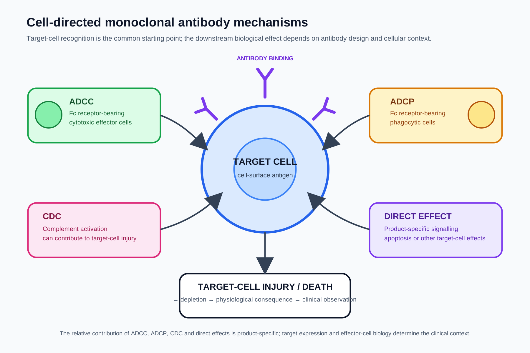
Figure 1. Cell-directed monoclonal antibody mechanisms. Antibody binding to a cell-surface antigen can recruit natural-killer-cell or myeloid effector functions, activate complement, or contribute to direct target-cell signalling or death. The clinical consequence depends on which normal and diseased cells express the target and on the duration and depth of depletion.
Effector Function as Part of the Pharmacological Mechanism
For antibodies whose therapeutic effect depends materially on immune-mediated depletion, the Fc region is part of the pharmacological design rather than merely a structural component. Fc interaction with Fc gamma receptors can recruit immune effector cells, while Fc interaction with complement components can contribute to complement-dependent cytotoxicity. The magnitude of these effects is influenced by antibody subclass, Fc glycosylation, engineering, target density and the biological environment.
The patient also contributes to the mechanism. The availability and functional state of natural killer cells, macrophages, neutrophils and complement components can affect how efficiently antibody-coated cells are removed. Disease state and concomitant treatment may therefore alter the observed pharmacodynamic response without changing the molecular identity of the product.
This patient-product interaction is especially relevant to pharmacovigilance because it prevents a simplistic interpretation of depletion-related events. A low blood-cell count, for example, may be consistent with the intended mechanism, but its clinical significance depends on the lineage affected, baseline reserve, duration, recovery and associated infection or other complications.
Fc-Mediated Effector Mechanisms
Three effector pathways are particularly useful for understanding cell-depleting antibodies: antibody-dependent cellular cytotoxicity (ADCC), antibody-dependent cellular phagocytosis (ADCP) and complement-dependent cytotoxicity (CDC). They are related but biologically distinct. Their relative contribution varies between products and target-cell systems, so the terms should not be treated as interchangeable descriptions of one process.
In ADCC, antibody-coated target cells are recognised by Fc receptors on cytotoxic immune effector cells, particularly natural killer cells. Engagement can trigger effector-cell activation and target-cell killing. ADCP uses Fc receptor-bearing phagocytes, such as macrophages, to recognise and ingest antibody-opsonised target cells. CDC involves activation of the complement system following antibody binding and can result in membrane injury and target-cell lysis. Other mechanisms, including direct induction of apoptosis or changes in target-cell signalling, may contribute depending on the antibody and antigen.
The distinction matters because each pathway has different biological prerequisites. A product that relies heavily on Fc receptor engagement may be affected by target-cell accessibility and the functional capacity of effector cells. A product for which complement activation is an important mechanism may have a different relationship between antigen density, antibody binding and cell killing. These mechanistic differences can influence both efficacy and adverse-event interpretation.
The Fc region can also be deliberately engineered. Changes to Fc sequence or glycosylation may increase, reduce or otherwise alter interactions with Fc receptors or complement. Such modifications are part of the product's molecular design and may therefore change the expected pharmacodynamic and safety profile. Pharmacovigilance should use the product-specific evidence rather than assuming that all IgG antibodies have equivalent effector activity.
Target Expression, Tissue Distribution and Selectivity
The safety profile of a cell-directed antibody begins with the distribution of its target antigen. A target that is largely restricted to diseased cells creates a different therapeutic window from one that is shared by the diseased population and an essential normal cell lineage.
Rituximab demonstrates this principle clearly. CD20 is expressed on B lymphocytes, including malignant B cells in the diseases for which rituximab is used. Depletion of normal B cells is therefore an expected pharmacological consequence as well as a therapeutic mechanism. EMA describes rituximab as binding CD20 and causing B-cell death, with therapeutic benefit in B-cell malignancies and several inflammatory conditions. [1]
Daratumumab provides another example. CD38 is present at high levels on abnormal plasma cells in multiple myeloma and AL amyloidosis, and EMA describes daratumumab binding CD38 and activating the immune system to kill abnormal white blood cells. Because CD38 is not exclusively restricted to the pathological cell population, the complete safety interpretation still depends on the extent and consequences of target engagement in normal tissues and cells. [2]
The relevant concept is therefore functional selectivity, not simply antigen specificity. An antibody can be highly specific for its molecular target while still producing predictable effects in normal cells that express that target. Pharmacovigilance assessment should map the target across relevant tissues and cell populations before deciding whether an event is plausibly on-target.
Depletion Versus Functional Modulation
Cell-surface binding does not by itself establish depletion. Some antibodies bind a cell and alter its function without causing substantial cell death. Others produce both functional modulation and depletion. The distinction should be maintained because the expected time course and safety consequences differ.
For a depleting antibody, pharmacodynamic assessment commonly focuses on the magnitude and duration of the affected-cell reduction and the subsequent recovery. For a function-modulating antibody, the relevant marker may instead be receptor occupancy, cellular activation, cytokine production or another measure of altered function. Where both mechanisms operate, the pharmacovigilance framework should recognise both pathways.
This distinction is particularly important when reviewing adverse events. A prolonged absence of a target-cell population may create delayed consequences that would not be predicted from the administration-day reaction alone. Conversely, an acute event may occur before measurable depletion is complete because antibody binding and immune activation can precede the full pharmacodynamic effect.
Representative Cell-Directed Antibodies
The following examples illustrate different target-cell contexts rather than defining a universal class safety profile.
| Product | Target | Principal cell context | Mechanistic illustration |
|---|---|---|---|
| Rituximab | CD20 | B lymphocytes, including malignant B cells | Antibody binding causes B-cell death; immune effector mechanisms contribute to depletion |
| Daratumumab | CD38 | Abnormal plasma cells and other CD38-expressing cells | Immune activation following CD38 binding contributes to killing of abnormal cells |
| Alemtuzumab | CD52 | Lymphocytes and other CD52-expressing immune cells | Targeting CD52 produces marked lymphocyte depletion |
| Ofatumumab | CD20 | B lymphocytes | CD20-directed depletion in multiple sclerosis; product-specific route and exposure influence the pharmacodynamic pattern |
These examples also show why the indication must remain part of the mechanistic interpretation. Rituximab is used to deplete B cells in both malignant and immune-mediated diseases, while ofatumumab is used for relapsing forms of multiple sclerosis. Alemtuzumab is used in relapsing-remitting multiple sclerosis and targets CD52 on lymphocytes. Daratumumab is directed toward a different cell population and has a distinct disease and treatment context. EMA product information confirms the relevant targets and therapeutic contexts for these products. [1–4]
The table should therefore be read as a classification aid rather than a statement that the listed products share identical effector pathways or safety outcomes.
Pharmacokinetics and Pharmacodynamics of Cell Depletion
The pharmacokinetics of a cell-directed antibody determine systemic exposure, but pharmacodynamics determine whether and for how long the target-cell population is altered. The relationship can be complicated by target-mediated drug disposition, tissue distribution, internalisation, binding to circulating cells and changes in the size of the target compartment during treatment.
When a substantial circulating target population is present, initial doses may encounter a large antigen sink. Binding and removal of target cells can change the disposition of subsequent doses because the target burden has changed. This can create nonlinear relationships between dose, serum concentration and pharmacodynamic effect. The interpretation of exposure-response relationships should therefore account for target dynamics rather than treating the antibody like a conventional small molecule with fixed distribution.
For depletion, a useful pharmacodynamic sequence is:
dose and exposure → target engagement → cell-count reduction → nadir → recovery → reconstitution of function
The timing of each stage can be clinically relevant. A safety event occurring during profound depletion should be interpreted differently from the same event occurring after substantial immune-cell recovery. Long-lived effects may also persist after measurable serum concentrations fall because the affected cell population or downstream physiological system may recover slowly.
Depth, Duration and Recovery of Depletion
The clinical significance of depletion depends on more than whether cells are removed. Depth describes the magnitude of the reduction; duration describes how long the population remains reduced; and recovery describes the return toward baseline or functional sufficiency. These dimensions can differ substantially between products, dosing schedules and patient populations.
Recovery may occur through production of new cells, maturation of precursor populations, redistribution from tissues, or other biological processes. The speed of recovery is therefore determined by the biology of the depleted lineage as well as the persistence of the antibody. A long serum half-life does not necessarily imply a proportionally long depletion period, and a declining serum concentration does not necessarily mean that the biological consequence has ended.
For pharmacovigilance, this creates a temporal framework for evaluating infections, cytopenias and other events. The relevant exposure window may extend beyond the administration date and beyond the period of measurable circulating antibody. Product-specific clinical data, pharmacodynamic markers and the known biology of the target population should be considered together.
Pharmacovigilance Consequences of Target-Cell Depletion
Once depletion is recognised as the pharmacodynamic mechanism, the safety assessment can be organised around the function of the affected cell population. This is more informative than treating depletion as an adverse event in itself. Depletion of a pathogenic or malignant population may be the desired therapeutic action; depletion of the corresponding normal population may be an expected pharmacological effect that becomes clinically important when it exceeds the patient's capacity to compensate.
B-cell-directed therapies illustrate this principle. Loss of B cells can reduce pathogenic antibody production and thereby contribute to therapeutic benefit, but sustained reduction of B-cell function can also influence humoral immune competence. The resulting infection risk, immunoglobulin changes and vaccine-response considerations are not interchangeable endpoints and should be evaluated according to the individual product, indication, patient population and observed evidence.
Lymphocyte-directed therapies can create a different pattern. Profound depletion can affect multiple components of immune surveillance and may be associated with delayed immune-mediated consequences. Alemtuzumab is a useful example of why a target-cell map matters: its target, CD52, is expressed on lymphocytes, and its pharmacological effect therefore extends beyond a single pathological cell population. EMA identifies alemtuzumab as a CD52-directed monoclonal antibody used in relapsing-remitting multiple sclerosis. [3]
In oncology, rapid destruction of a substantial malignant cell burden can produce acute metabolic consequences. Such events are not simply generic "immune reactions"; they can arise from the biological consequences of extensive target-cell killing. Conversely, infusion-related symptoms can reflect acute immune activation, cytokine release or other administration-associated mechanisms and should not automatically be attributed to the same pathway as delayed depletion-related events.
The mechanistic framework is therefore:
target expression → degree of depletion → physiological function lost or altered → time to consequence → clinical event
The chain provides a structured hypothesis for case assessment while preserving the distinction between pharmacological plausibility and demonstrated causality.
Acute Events During Target-Cell Killing
Cell-depleting antibodies can produce acute reactions around administration, particularly when substantial numbers of target cells are present or when immune effector mechanisms are rapidly activated. The clinical phenotype may include fever, chills, hypotension, respiratory symptoms or other systemic manifestations, but the mechanism must be assessed product by product.
The timing of the event is informative but not definitive. An event beginning during or shortly after infusion is compatible with an acute administration-related mechanism, whereas an event developing later may be more consistent with the downstream consequences of depletion. Concomitant medicines, underlying disease and the clinical setting must also be considered.
In case processing, the suspect medicinal product should remain clearly distinguished from concomitant therapies and from the underlying disease. A cell-depleting mechanism can provide biological context for the event, but it does not establish that the event is caused by the product. This is particularly important in oncology, where disease progression and combination treatment can generate overlapping clinical manifestations.
Infection and Immune Consequences
Infection assessment after cell depletion requires a time-dependent approach. The relevant question is not simply whether an infection occurred after treatment, but whether the depth and duration of the affected immune-cell reduction provide a credible biological context for the event and whether other risk factors are present.
Potential contributors include the underlying disease, previous or concomitant immunosuppressive treatment, baseline immune status, age, comorbidity, prior infections and the degree of immune-cell recovery. The product's route, dose and treatment schedule may also influence the pharmacodynamic exposure.
For signal evaluation, these variables help distinguish a plausible mechanism from a crude temporal association. A cluster of infections occurring during a period of sustained target-cell depletion may merit a different assessment from isolated infections occurring without measurable pharmacodynamic suppression. The appropriate analysis should use the evidence available for the product rather than applying a universal threshold to all cell-depleting antibodies.
Cytopenias, Immunoglobulins and Other Biomarkers
Laboratory abnormalities can provide important pharmacodynamic evidence when interpreting safety reports. Cell counts can demonstrate depletion or recovery, while immunoglobulin concentrations may provide information about the functional consequences of B-cell-directed treatment. Such markers should not automatically be treated as adverse reactions: some are expected pharmacodynamic effects, whereas others become clinically significant depending on magnitude, persistence and associated symptoms or complications.
The distinction between a pharmacodynamic effect and an adverse reaction is particularly useful in periodic safety evaluation. A reduction in a target-cell population can be evidence that the product is exerting its intended mechanism. The same biological change may become a safety concern when it is excessive, prolonged, associated with serious clinical consequences, or occurs in a population for which the therapeutic window is narrower.
Immunogenicity and Loss of Pharmacological Effect
Monoclonal antibodies can induce anti-drug antibodies (ADAs), and the clinical significance of immunogenicity is product- and patient-dependent. EMA's current effective guideline on immunogenicity assessment of therapeutic proteins recommends an integrated assessment using immunological, pharmacokinetic, pharmacodynamic, efficacy and safety information. The monoclonal-antibody-specific guideline is an addendum addressing unwanted immunogenicity of monoclonal antibodies intended for in-vivo clinical use. [5–6]
For a cell-depleting antibody, ADAs may potentially alter exposure, target engagement or persistence of the pharmacodynamic effect. Whether that occurs, and whether it affects safety or efficacy, must be demonstrated for the individual product. The absence of an observed pharmacodynamic response should therefore not automatically be attributed to immunogenicity; alternative explanations include target-cell characteristics, inadequate exposure, disease biology, concomitant treatment and assay limitations.
An integrated immunogenicity assessment is consequently more informative than simply reporting ADA frequency. Where available, ADA status should be interpreted alongside serum concentrations, pharmacodynamic markers, clinical response and adverse events.
Signal Detection and Signal Evaluation
Cell-directed mechanisms can improve signal detection because they provide a biological hypothesis linking product exposure to a defined physiological consequence. However, mechanistic plausibility is only one component of signal evaluation. The observed clinical pattern, alternative explanations, reporting characteristics, epidemiology and evidence from other data sources remain necessary.
A useful evaluation asks whether the signal follows the expected pharmacological sequence. For example, if an adverse outcome is hypothesised to result from prolonged B-cell depletion, evidence of B-cell suppression or impaired immune function during the relevant exposure period may strengthen the hypothesis. Conversely, if the event occurs without meaningful target-cell engagement or during a period of recovery, the proposed mechanism may be less persuasive.
Signal evaluation should also distinguish class effects from product-specific effects. Products directed at the same antigen may differ in epitope, affinity, Fc activity, dose, route, exposure and treatment population. Biosimilars and reference products require particular attention to product identification and traceability when evaluating product-specific safety information. EMA's GVP guidance for biological medicinal products identifies continuous product and batch traceability as a key pharmacovigilance requirement. [7]
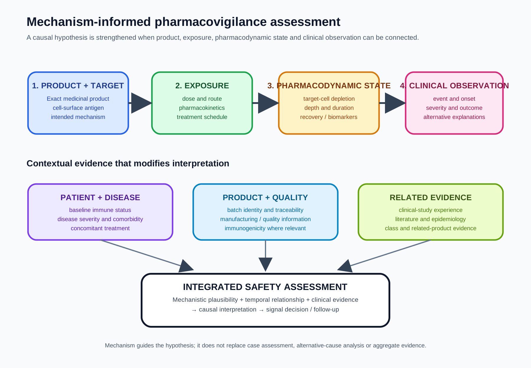
Figure 2. Mechanism-informed assessment of a cell-directed antibody. Product and target characteristics establish the mechanistic hypothesis; exposure and depletion determine the pharmacodynamic context; the resulting biological consequences provide the bridge to clinical observation. Patient, disease, product-quality and related-product information modify interpretation rather than replacing the core causal pathway.
Class Effects and Product-Specific Effects
The term "cell-depleting antibody" describes a mechanism, not a complete safety phenotype. Two products can target the same cell lineage while producing different degrees of depletion because of differences in antigen recognition, Fc-mediated activity, dose, route, exposure or treatment schedule. Conversely, products directed at different antigens may produce overlapping clinical consequences if they deplete cells with similar physiological functions.
This distinction is important when using class experience in signal assessment. Class knowledge can provide a biologically reasonable hypothesis, but product-specific evidence determines whether the hypothesis applies to the medicinal product under review. A safety signal should therefore not be transferred mechanically from one antibody to another solely because both are described as cell-depleting.
Practical Pharmacovigilance Implementation
The mechanism of a cell-directed antibody becomes operationally useful only when it is connected to the records and processes through which pharmacovigilance is performed. The objective is not to label every event as "on-target", but to preserve enough information to test the mechanistic hypothesis during individual case assessment, aggregate review and signal evaluation.
At case level, useful information may include the exact medicinal product and formulation, indication, dose and administration date, concomitant treatment, baseline and subsequent target-cell measurements, relevant laboratory findings, clinical onset, treatment of the event and outcome. Where product or batch information is available, it should be retained in accordance with the applicable pharmacovigilance process. For biological medicinal products, EMA specifically emphasises continuous product and batch traceability so that product-specific safety and immunogenicity concerns can be detected and evaluated. [7]
At aggregate level, the same information allows events to be analysed against exposure and pharmacodynamic state. Counts of adverse events without the corresponding patient population, treatment duration, target-cell depletion or recovery information can obscure the relationship between exposure and biological effect. The quality of the analysis therefore depends partly on whether the pharmacovigilance system captures the variables needed to test the mechanism.
Roles and Interfaces
The pharmacovigilance function does not generate all of the evidence needed to interpret a cell-depleting mechanism. Effective assessment can require interfaces with clinical safety, clinical pharmacology, immunogenicity, medical affairs, regulatory, quality and manufacturing functions.
Clinical and medical teams may provide information about the target disease, treatment patterns and differential diagnosis. Clinical pharmacology can help interpret exposure and pharmacodynamic relationships. Immunogenicity specialists can assess ADA findings and assay limitations. Quality and manufacturing functions may be required when a potential safety pattern appears associated with a product or batch characteristic. Pharmacovigilance integrates these inputs into the safety assessment and ensures that conclusions are appropriately documented and governed within the pharmacovigilance system.
This division of responsibility is consistent with the general GVP principle that pharmacovigilance activities operate through a quality-assured system of structures, processes and outcomes. GVP Module I distinguishes legal requirements from implementation guidance: applicable legal requirements are expressed using "shall", while implementation guidance uses "should". [8]
Evidence and Traceability
A mechanism-informed assessment should be reproducible. The reviewer should be able to identify which product was involved, what target was relevant, what evidence established depletion or other cellular effects, what clinical event occurred, and how alternative explanations were considered.
For individual case safety reports, the information required by GVP Module VI concerns the collection, management and submission of suspected adverse-reaction reports. The module distinguishes adverse reactions from patterns of use that do not themselves constitute suspected adverse reactions, although such information may remain relevant to aggregate safety evaluation. [9]
For cell-directed antibodies, the practical implication is that mechanistic information should be preserved without allowing the mechanism to substitute for case validity or clinical assessment. A well-documented pharmacodynamic observation can strengthen a causal hypothesis; it does not remove the need to assess seriousness, expectedness, alternative causes, dechallenge or rechallenge where applicable, and the overall clinical course.
Common Analytical Failure Modes
Several errors recur when a cell-depletion mechanism is interpreted without sufficient biological context.
Treating target expression as proof of causality. A target can be expressed on a cell without the reported event being caused by depletion of that cell. The mechanism establishes plausibility, not certainty.
Equating depletion with an adverse reaction. Removal of the target population may be the intended pharmacological action. Safety significance depends on depth, duration, physiological consequences and clinical context.
Ignoring recovery kinetics. An adverse event may arise while the target population remains suppressed even after serum drug concentrations have declined. Conversely, an event occurring after recovery may require a different explanation.
Assuming identical mechanisms within an antigen class. Antibodies directed at the same antigen can differ in epitope, Fc activity, dose, route and exposure. These differences can change the biological effect.
Confusing acute administration reactions with delayed depletion consequences. Both may follow treatment, but their timing and biological pathways can differ.
Over-attributing infection risk to the antibody. Infection susceptibility is influenced by disease, concomitant immunosuppression, prior therapy, patient factors and the depth and duration of immune impairment.
Failing to distinguish product identity. For biologicals and particularly related products with the same active-substance name, inadequate product identification can prevent meaningful product-specific signal evaluation.
These are analytical failure modes rather than statements that a particular product or pharmacovigilance system has failed. Their value is as a framework for testing whether the evidence supports the proposed mechanism.
Inspection Perspective
An inspector evaluating pharmacovigilance for a cell-directed antibody could reasonably examine whether the system can demonstrate the connection between the known pharmacology and the safety activities performed. The focus should be on evidence and effectiveness rather than on whether a particular document contains a preferred phrase.
Illustrative inspection questions include:
| Area | Illustrative question |
|---|---|
| Product definition | Can the system distinguish the medicinal product from related biologicals and capture available batch information? |
| Mechanism | Is the target-cell mechanism documented and scientifically consistent with current product knowledge? |
| Case processing | Are clinically relevant depletion, laboratory and timing data captured when available? |
| Aggregate assessment | Are safety observations considered in relation to exposure and pharmacodynamic state? |
| Signal management | Can reviewers explain why a proposed signal is biologically plausible and what evidence could weaken the hypothesis? |
| Immunogenicity | Are ADA findings interpreted with pharmacokinetic, pharmacodynamic, efficacy and safety information where relevant? |
| Governance | Are conclusions, decisions, supporting evidence and follow-up actions traceable within the quality system? |
The purpose of such questions is to determine whether the pharmacovigilance system can detect and evaluate changes in the benefit-risk balance effectively. GVP Module I describes the pharmacovigilance system in terms of structures, processes and outcomes and requires quality systems that are adequate and effective for the performance of pharmacovigilance activities. [8]
Risk Management and Ongoing Benefit-Risk Evaluation
The mechanism of cell depletion can inform risk management because it identifies predictable pathways from target engagement to clinical consequence. It can therefore help define which risks or important potential risks deserve particular attention, what evidence is needed to characterise them and which pharmacodynamic or clinical measures can contribute to ongoing evaluation.
Risk management remains product-specific. GVP Module V provides the framework for risk management systems, while the product's clinical evidence and evolving post-authorisation experience determine which risks require specific management. A mechanistic class description should not be converted automatically into a product-specific safety concern without supporting evidence.
The same principle applies to periodic safety evaluation and signal management. A plausible mechanism can guide the search for relevant evidence, but the conclusion should integrate individual cases, aggregate data, epidemiology where available, clinical studies, literature, non-clinical information and product-quality information as appropriate.
A Practical Assessment Framework
When assessing a suspected safety issue involving a cell-directed monoclonal antibody, the following sequence provides a disciplined starting point:
- Identify the product and target. Confirm the exact medicinal product and the cell-surface antigen involved.
- Define the intended mechanism. Determine whether the product is intended to deplete cells, modulate their function, or combine mechanisms.
- Map target expression. Identify the relevant diseased and normal cell populations and the physiological functions they serve.
- Characterise exposure. Consider dose, route, treatment schedule, pharmacokinetics and factors affecting target-mediated disposition where relevant.
- Establish pharmacodynamic state. Review target-cell counts, depletion depth, duration, recovery and other validated biomarkers when available.
- Place the event in time. Relate onset and course to administration, depletion and recovery rather than relying on exposure alone.
- Assess alternative explanations. Consider disease, concomitant medicines, baseline status, infections, procedures and other relevant causes.
- Integrate immunogenicity where relevant. Assess ADAs together with exposure, pharmacodynamics, efficacy and safety rather than in isolation.
- Assess product specificity. Determine whether the evidence supports a class effect, a product-specific effect, or remains insufficient to distinguish them.
- Document the reasoning. Preserve the evidence, uncertainty, decision and follow-up so that the assessment can be reproduced and revisited.
This sequence keeps mechanism at the centre of the assessment without allowing mechanistic plausibility to replace clinical evidence.
Key Takeaways
Cell-directed monoclonal antibodies are defined by their recognition of cell-surface targets and the biological consequences of that recognition. Many important products produce therapeutic effects through target-cell depletion, but depletion is not a single mechanism: Fc-mediated cytotoxicity, phagocytosis, complement activation and direct cellular effects can contribute in different proportions.
For pharmacovigilance, the critical conceptual bridge is target → engagement → depletion or modulation → physiological consequence → clinical observation. Target expression determines what can be affected; antibody design and exposure influence the magnitude of the effect; and the depth and duration of depletion determine when downstream consequences may appear.
The class therefore cannot be assigned a single universal safety profile. Rituximab, daratumumab, alemtuzumab and ofatumumab illustrate different target-cell populations and clinical contexts. Product-specific evidence remains necessary, while class biology provides a structured hypothesis for assessment.
The most useful pharmacovigilance approach is consequently mechanism-informed but evidence-led: preserve product and batch identity, connect cases and aggregate data to pharmacodynamic state where possible, integrate immunogenicity and exposure information, distinguish expected pharmacology from clinically significant adverse reactions, and document the reasoning sufficiently for effective oversight.
References and Regulatory Sources
- European Medicines Agency. MabThera (rituximab): EPAR – Medicine overview and product information. EMA. Current product information accessed September 2026. https://www.ema.europa.eu/en/medicines/human/EPAR/mabthera
- European Medicines Agency. Darzalex (daratumumab): EPAR – Medicine overview, risk management plan and product information. EMA. Current product information accessed September 2026. https://www.ema.europa.eu/en/medicines/human/EPAR/darzalex
- European Medicines Agency. Lemtrada (alemtuzumab): EPAR – Medicine overview and product information. EMA. Current product information accessed September 2026. https://www.ema.europa.eu/en/medicines/human/EPAR/lemtrada
- European Medicines Agency. Kesimpta (ofatumumab): EPAR – Medicine overview and product information. EMA. Current product information accessed September 2026. https://www.ema.europa.eu/en/medicines/human/EPAR/kesimpta
- European Medicines Agency. Guideline on immunogenicity assessment of therapeutic proteins – Revision 1. EMEA/CHMP/BMWP/14327/2006 Rev. 1. Legal effective date 1 December 2017. https://www.ema.europa.eu/en/immunogenicity-assessment-biotechnology-derived-therapeutic-proteins-scientific-guideline
- European Medicines Agency. Guideline on immunogenicity assessment of monoclonal antibodies intended for in-vivo clinical use. EMA/CHMP/BMWP/86289/2010. https://www.ema.europa.eu/en/immunogenicity-assessment-monoclonal-antibodies-intended-vivo-clinical-use-scientific-guideline
- European Medicines Agency. GVP Product- or Population-Specific Considerations II: Biological medicinal products. Section P.II.A.1.4, Product traceability. https://www.ema.europa.eu/en/documents/scientific-guideline/guideline-good-pharmacovigilance-practices-gvp-product-or-population-specific-considerations-ii-biological-medicinal-products_en.pdf
- European Medicines Agency. GVP Module I – Pharmacovigilance systems and their quality systems. EMA/541760/2011. https://www.ema.europa.eu/en/documents/scientific-guideline/guideline-good-pharmacovigilance-practices-module-i-pharmacovigilance-systems-and-their-quality-systems_en.pdf
- European Medicines Agency. GVP Module VI – Collection, management and submission of reports of suspected adverse reactions to medicinal products. Rev. 2, EMA/873138/2011. https://www.ema.europa.eu/en/documents/scientific-guideline/guideline-good-pharmacovigilance-practices-gvp-module-vi-collection-management-submission-reports-suspected-adverse-reactions-medicinal-products-tracked-changes-rev-2_en.pdf
Regulatory Note
This article distinguishes legally binding EU pharmacovigilance requirements from GVP guidance and from recommended scientific or operational practice. The GVP Modules cited above provide guidance for implementation of the EU pharmacovigilance framework; they should not be read as creating requirements beyond the applicable legislation. Product-specific scientific and safety conclusions should be based on the current authorised product information and relevant regulatory assessment rather than on the mechanism class alone.